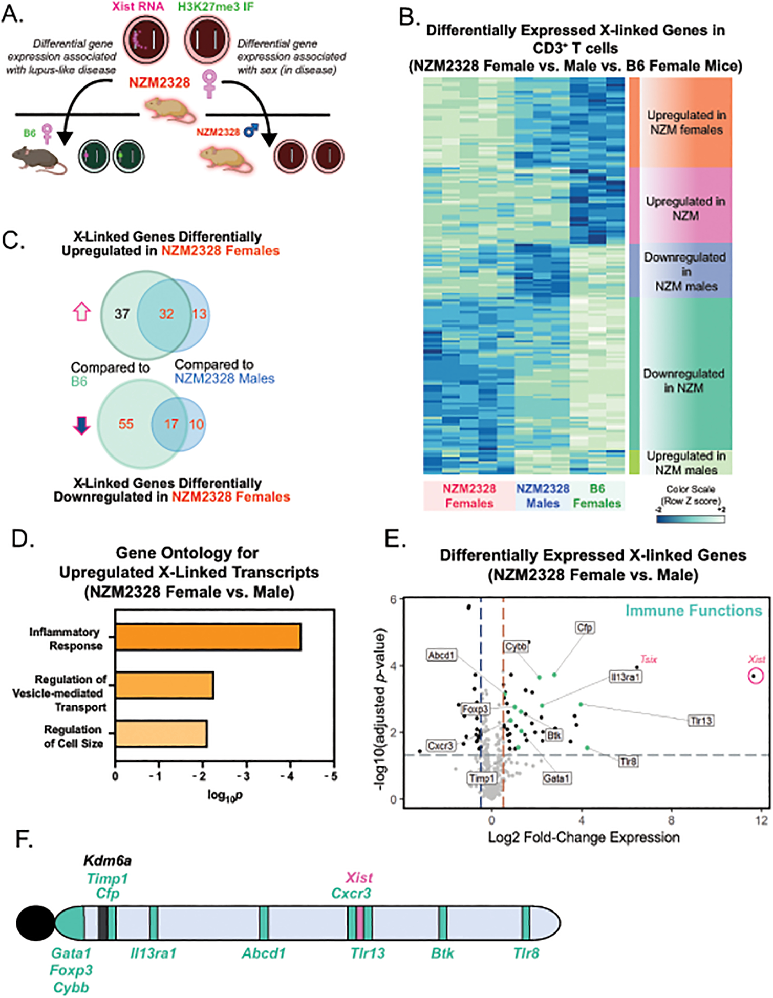
Figure 5. Activated T cells from NZM2328 mice exhibit female-biased expression of X-linked genes with immune functions.

A. Schematic for the experimental approach to determine sex-specific disease expression profiles using in vitro activated T cells from 18–24-week old NZM2328 male (n=3), NZM2328 female (n=5), and B6 female (n=3) mice. B. Heatmap of differentially expressed X-linked genes for in vitro activated T cells from NZM2328 female and male mice and B6 female mice. C. Venn-diagrams showing the number of differentially upregulated and downregulated X-linked genes in in vitro activated T cells from NZM2328 female mice relative to B6 female or NZM2328 male mice. D. Gene Ontology (GO) analyses for Biological Processes of upregulated X-linked genes in in vitro activated T cells comparing NZM2328 female and NZM2328 male mice. E. Volcano plot of differentially expressed X-linked genes in activated T cells from NZM2328 female relative to age-matched NZM2328 male mice. X-linked genes with known immune functions are colored in teal. F. Location of the differentially upregulated immune-related X-linked genes across the X chromosome. The known XCI escape gene, Kdm6a, which is also overexpressed in NZM2328 females, is also shown in black.
