
Figure 7.
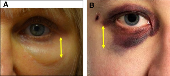
The barrier function of the malar septum. The left image demonstrates periocular ecchymosis and the right image is a closeup of the post-hyaluronic acid recurrent eyelid edema patient described in Case 4. The yellow arrows demonstrate the inferolateral portion of the malar septum. Note the stark transition between (A) ecchymosis and normal tissue and the (B) stark transition between edema and normal tissue (right).