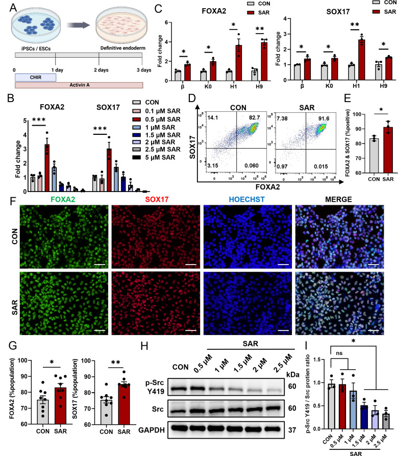
Fig. 1.
Low concentrations of SAR promote Definitive Endoderm (DE) differentiation. DE differentiation was performed using the previously reported protocol (A) for 3 days. After 3 days of differentiation, total RNAs were extracted and the expressions of FOXA2 and SOX17 in DE cells derived from the H1 cells were determined by qPCR (B). (C) QPCR results of FOXA2 and SOX17 in two different ESC lines (H1 and H9) and two iPSC lines (K0 and β) treated with 0.5 µM SAR. (D) Flow cytometric analysis of DE cells derived from hESC line H1 expressing FOXA2 and SOX17 with or without the treatment of 0.5 µM SAR. (E) Quantitative statistics of FOXA2+/SOX17+ cells corresponding to (D). (F) Immunofluorescent staining examined FOXA2+ (green) and SOX17+ (red) cells treated with 0.5 µM SAR differentiated from hESC line H1. The nucleus was counterstained with Hoechst 33,342. Quantitative statistics were shown in (G). (H) The p-Src Y419 and Src protein levels in DE cells treated with different concentrations of SAR were detected using western blotting. GAPDH was used as a loading control. (I) Quantitative statistics of p-Src Y419/Src corresponding to H. Data are presented as mean ± SEM (n = 3–8). *p < 0.05; **p < 0.01; ***p < 0.001; ns, not significant

