Fig. 5.
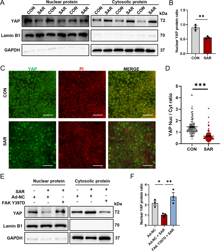
SAR inhibits YAP nuclear translocation. Human ESC line H1 was differentiated into DE cells with or without the addition of SAR. Three days after differentiation, nuclear protein and cytosolic protein were isolated separately and YAP protein level was determined by western Blotting (A). In the nuclear fraction, Lamin B1 was used as loading control and GAPDH as negative control; in the cytosolic fraction, GAPDH was used as loading control and Lamin B1 as negative control. (B) Quantitative statistics of YAP corresponding to (A). (C) Immunofluorescent staining examines the subcellular location of YAP (green) with or without 0.5 µM SAR treatment. The nucleus was counterstained with PI. Scale bars = 50 μm. The YAP fluorescent intensity within nucleus or cytoplasm was measured separately using ImageJ and the nuclear-to-cytoplasmic ratio is shown in (D). N = 118 for control group, n = 108 for SAR-treated group. (E) Western blotting for YAP in DE cells treated with negative control adenovirus (Ad-NC) or FAK Y397D-overexpressing adenovirus, in conjunction with either DMSO or SAR during 3 days of DE differentiation. In the nuclear fraction, Lamin B1 was used as loading control and GAPDH as negative control; in the cytosolic fraction, GAPDH was used as loading control and Lamin B1 as negative control. (F) Quantitative statistics of YAP corresponding to (E). Data are presented as mean ± SEM (n = 3). *p < 0.05; **p < 0.01; ***p < 0.001

