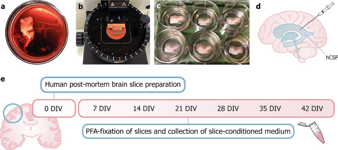
Fig. 1.
Preparation of HPMB-OSCs. (a) A tissue block of the middle frontal gyrus is dissected during autopsy and cut into a block of roughly 1.5 × 1.5 cm. (b) A vibratome is used to cut 300 μm-thick slices. (c) Slices are transferred onto semi-permeable membrane inserts in 6-well culture plates containing slice culture medium. (d) hCSF is collected at autopsy from the lateral ventricles using a syringe and spinal needle and slices are cultured with or without hCSF supplementation to the culture medium. (e) Timeline of the experimental procedure: slices are prepared and cultured up to 42 DIV. Slices are fixed in PFA once every week and slice-conditioned medium is collected for further analysis

