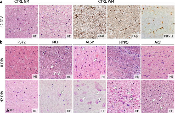
Fig. 2.
HPMB-OSCs maintain relatively normal tissue structure and diversity of neural cell types. (a) Control HPMB-OSCs show preservation of gross tissue structure based on HE staining and presence of neurons in the grey matter (GM) and GFAP+ astrocytes, Olig2+ oligodendrocytes and P2RY12+ microglia in the white matter (WM) at 42 DIV. Expression of P2RY12, a marker for non-activated microglia, together with the ramified morphology, indicates a homeostatic cell state of microglia in HPMB-OSCs even up till 42 DIV. (b) HE staining of uncultured reference slices (0 DIV) and slices cultured without hCSF (42 DIV) from multiple donors shows relative preservation of the histo-architecture, but a clear reduction in total cell numbers, after six weeks in culture. Additionally, disease-specific pathology is observed in both reference and cultured slices, as indicated by white arrows. This includes enlarged, rounded microglia/macrophages in MLD patient-derived slices in the white matter and at the subcortical boundary. Axonal spheroids (reference slice) and pigmented glia (cultured slice) are observed in ALSP patient slices and robust reactive astrogliosis in HYPO patient slices. Small Rosenthal fibers are observed in AxD patient slices at 42 DIV

