Fig. 3.
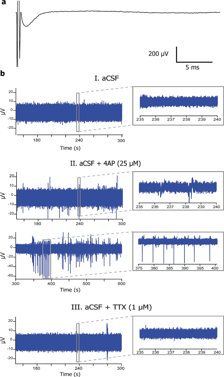
Electrophysiological recordings in HPMB-OSCs demonstrate neuronal activity ex vivo. (a) Field potential recorded upon electrical stimulation of a HPMB slice obtained from CTRL3 at 11 DIV shows neuronal activity ex vivo. (b) Representative examples of extracellular activity recorded from a HPMB slice obtained from the MS patient at 11 DIV during the incubation with normal aCSF (panel I), aCSF with 25 µM 4AP (panel II), and aCSF with 1 µM TTX (panel III). 4AP increases neuronal activity, whereas TTX abolishes the neuronal firing. Inserts show an expanded view of the recorded events at the time period indicated by the box

