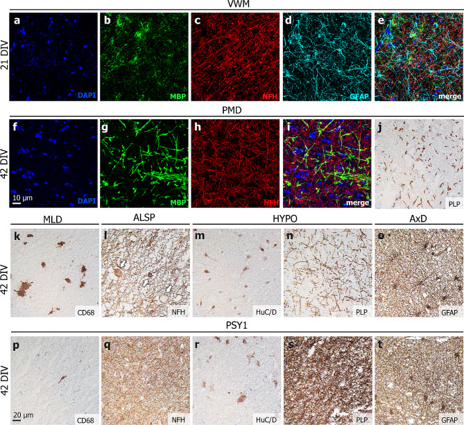
Fig. 4.
Disease-specific neuropathology in leukodystrophy patient-derived HPMB-OSCs. (a-e) Free-floating staining of whole-mount slices of the VWM patient at 21 DIV shows paucity of MBP+ myelin, relative preservation of NFH+ axons and dysmorphic GFAP+ astrocytes. (f-i) Free-floating staining of whole-mount PMD patient-derived slices at 42 DIV shows a lack of MBP+ myelin, (j) which is also appreciated when staining paraffin-embedded sections for PLP. (k) In MLD patient slices, CD68+ microglia/macrophages show increased lysosomal activity. (l) ALSP patient-derived HPMB-OSC demonstrates NFH+ axonal spheroids of different sizes (white arrows). (m) In HYPO patient slices, altered staining patterns for neuronal marker HuC/D and (n) hypomyelination on PLP staining can be appreciated. (o) AxD patient slices show dysmorphic, double-nucleated GFAP+ astrocytes. (p-t) None of the described neuropathological features are observed in HPMB-OSCs derived from control or psychiatric donors

