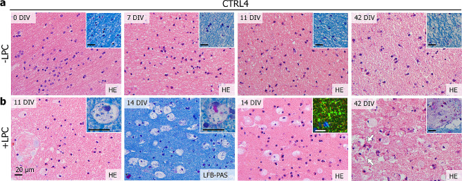
Fig. 6.
Lysolecithin treatment of HPMB-OSCs induces a multicellular response to injury ex vivo. (a) HPMB-OSCs obtained from CTRL4 that are not treated with LPC show normal tissue structure and cell morphology based on HE and LFB-PAS staining (inserts) at 0 DIV (uncultured reference slices) and 7, 11 and 42 DIV. (b) Slices treated with LPC show many rounded microglia/macrophages at 11, 14 and 42 DIV. The microglia/macrophages have an enlarged cytoplasm and show LFB+ vacuoles (11 DIV insert) and sometimes PAS+ vacuoles (14 DIV insert). At 14 DIV, myelin swelling is observed based on MBP (green) and NFH (red) staining (insert). At 42 DIV, rounded macrophages are still present, as well as signs of reactive astrogliosis (white arrows). The macrophages are also found close to the blood vessels and even inside the vascular adventitia (42 DIV insert). Data obtained using 1.0 mg/ml LPC are shown. A similar but milder response was obtained using 0.5 mg/ml LPC. All scale bars represent 20 μm

