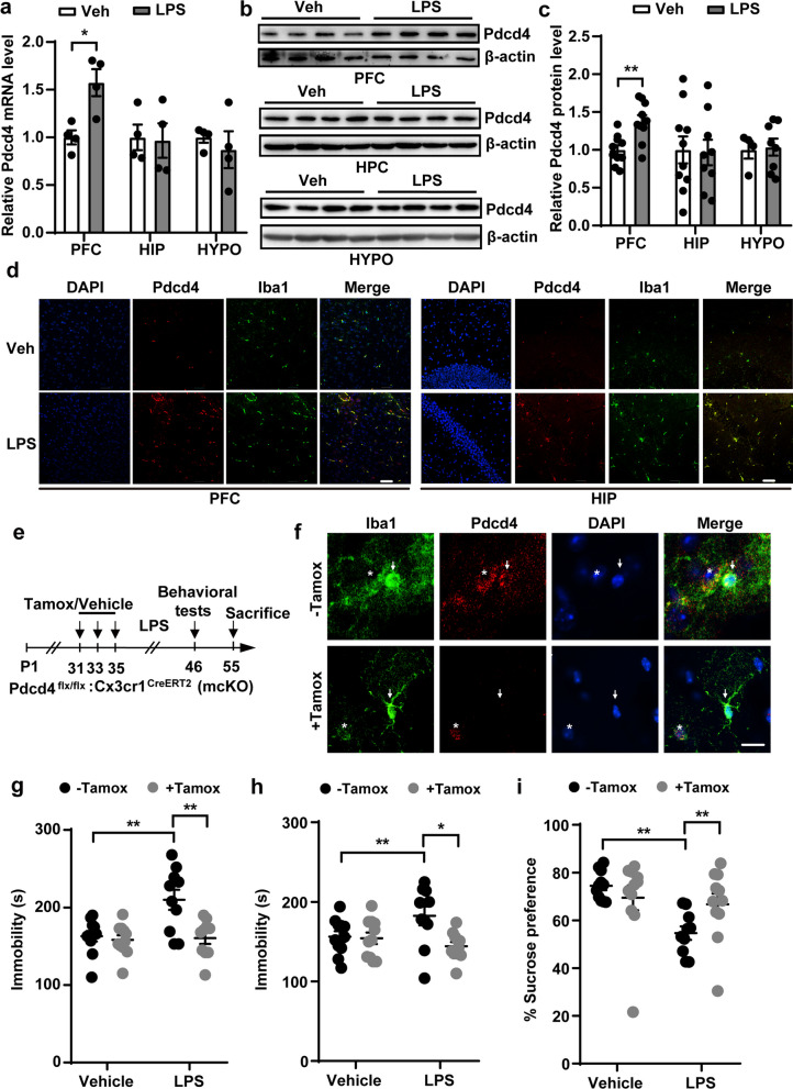
Fig. 1.
Microglial knockout of Pdcd4 ameliorates neuroinflammation related depression. a The change of mRNA levels of Pdcd4 in the prefrontal cortex (PFC), the hippocampal (HIP) and the hypothalamus (HYPO) after LPS administration for 10 days. Unpaired two-tailed Student’s t test, *P < 0.05. b, c The change of protein levels of Pdcd4 in the prefrontal cortex (PFC), the hippocampal (HIP) and the hypothalamus (HYPO) after LPS administration for 10 days. Unpaired two-tailed Student’s t test, **P < 0.01. d Iba1 antibody (Green), Pdcd4 antibody (Red) and DAPI (blue) was used into immunostaining in the PFC or HIP. N = 3 per group, scale bar = 50 μm. e Schematic diagram of animal experiments of LPS-induced mice. f Iba1 (Green) and Pdcd4 (Red) immunostaining in the PFC. Arrow represents microglia, star represents cell which is adjacent to and surrounded by microglia. N = 3 per group, scale bar = 20 μm. g Immobility time in TST (Veh vs. LPS: F1,39 = 8.259, P < 0.01; − Tam vs. + Tam: F1,39 = 9.949, P < 0.01), h immobility time in FST (Veh vs. LPS: F1,39 = 1.025, P = 0.317; − Tam vs. + Tam: F1,39 = 6.401, P < 0.01), and i sucrose consumption in SPT under basal or LPS conditions in the mcKO mice with or without Tamoxifen (Veh vs. LPS F1,39 = 8.506, P < 0.01; − Tam vs. + Tam F1,39 = 0.846, P = 0.363). Two-ways ANOVA and Sidak’s multiple comparisons test, *P < 0.05, **P < 0.01

