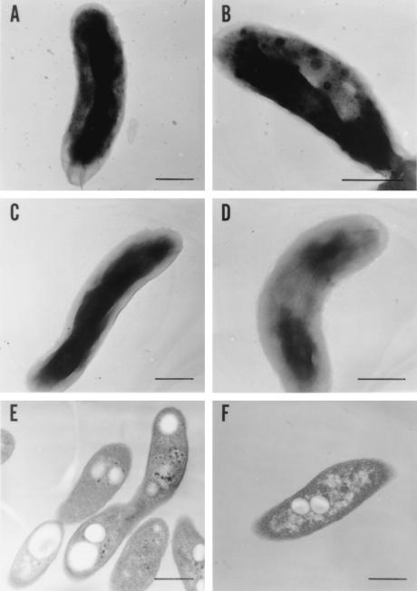
FIG. 3.
Electron micrographs of V. cholerae strains. The bacteria were harvested at 0 and 2 h after the addition of Pi, as for Fig. 2D. Bars, 0.5 μm. Negatively stained samples were prepared from the wild-type culture after 0 (C) and 2 (A and B) h of incubation and from KCV3 (the ppk mutant) after 2 h of incubation (D). Thin-section samples were prepared from the wild type (E) and KVC3 (F) after 2 h of incubation. Magnifications, ×28,000 (A, C, E, and F), ×35,000 (D), and ×45,000 (B).

