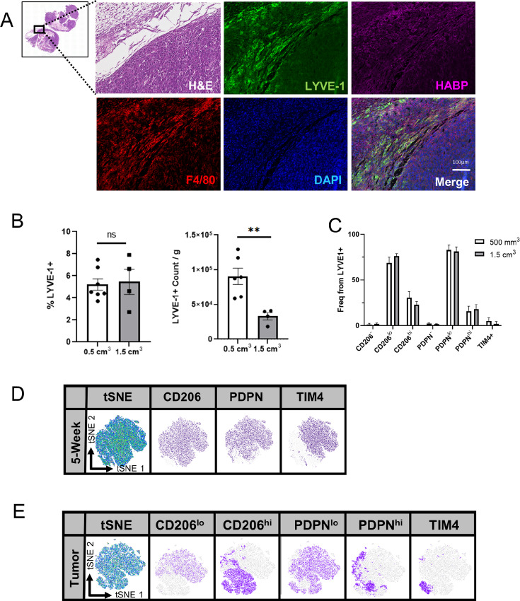
FIGURE 2.
Identification and localization of LYVE-1+ macrophages in mammary tumors. A, Representative images from formalin-fixed and paraffin-embedded (FFPE) sections of EO771 tumors from Csf1rfl/fl mice stained by H&E, and immunostained for LYVE-1 (green), HABP (magenta), F4/80 (red), and DAPI (blue; n = 5). CD45+F4/80+CD11b+LYVE-1+ macrophage count, CD45+F4/80+CD11b+LYVE-1+ macrophage count normalized to tumor weight (B), and frequency of CD206, PDPN, and TIM4 from CD45+F4/80+CD11b+LYVE-1+ macrophage subset in EO771 tumors from C57BL/6 mice (C). FlowJo generated tSNE plots from CD45+F4/80+CD11b+LYVE-1+ subset in 5-week female mammary glands (D), and EO771 C57BL/6 tumors (E). **, P < 0.01. Scale bars, 100 µm. Each dot represents one mouse.

