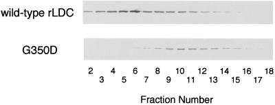
FIG. 4.
Analysis of wild-type rLDC and G350D mutant rLDC by gel filtration. Cells of E. coli DH5α harboring plasmid pTLDC or pG350D were grown in 4 ml of 2× TY medium containing ampicillin (100 μg/ml) at 37°C for 18 h and collected by centrifugation at 10,000 × g for 5 min at 4°C. The cells were suspended in 2 ml of dimer buffer (20 mM potassium phosphate [pH 6.7] containing 0.5 mM EDTA, 1 mM dithiothreitol, 0.1 M NaCl, 50 μM PLP, 0.5 mM ornithine) (33) and sonicated. The sonicated extracts were filtrated by column chromatography with a Superdex 200 HR 10/30 column (Pharmacia) equilibrated with dimer buffer. Proteins in the fractions were analyzed by SDS-PAGE and then transferred to nitrocellulose membrane (Hybond-C pure; Amersham), and the LDC protein was visualized immunologically using anti-LDC antibodies and anti-rabbit immunoglobulin G(Fc)-alkaline phosphatase conjugate (Seikagaku Kogyo Co., Tokyo, Japan).

