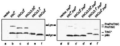
FIG. 4.
Western blot analysis (for conditions see Materials and Methods) of E. coli SCS1 cell extracts (2 μl/lane) in the absence (left) or the presence (right) of traF. Plasmids used are as follows. Lane a, pMS119EH (vector); lane b, pRE178 (trbC+); lane c, pRE178Δ3 (trbCΔ3+); lane d, pRE178Δ3.05 (trbCΔ3.05+); lane e, pRE178Δ3.1 (trbCΔ3.1+); lane f, pRE178Δ4 (trbCΔ4+); lane a′, pJH472 (traF+) and pMS119EH (vector); lane b′, pJH472 (traF+) and pRE178 (trbC+); lane c′, pJH472 (traF+) and pRE178Δ3 (trbCΔ3+); lane d′, pJH472 (traF+) and pRE178Δ3.05 (trbCΔ3.05+); lane e′, pJH472 (traF+) and pRE178Δ3.1 (trbCΔ3.1+); lane f′, pJH472 (traF+) and pRE178Δ4 (trbCΔ4+). Standard molecular mass markers (Rainbow labeled markers, low range; Amersham Pharmacia Biotech): Lys, lysozyme (14.3 kDa), and Apr, aprotinin (6.5 kDa). Positions of the 145-aa PreProTrbC, N-terminally cleaved ProTrbC, N- and C-terminally processed TrbC∗, and (circular) pilin (TrbC) are indicated on the right side of the figure.

