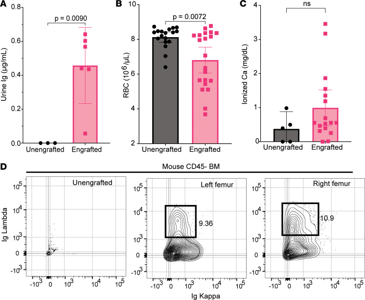
Figure 5. Myeloma-engrafted NSG+hIL6 mice with sequelae of human disease.
(A) Urine from NSG+hIL6 mice (n = 6) engrafted 15 weeks previously with MM1 BM cells and unengrafted controls (n = 3) was evaluated for human Ig. (B) RBC counts from engrafted (n = 18) versus unengrafted (n = 21) mice at 15 weeks after injection. (C) Serum ionized calcium concentrations in engrafted (n = 15) compared with unengrafted controls (n = 5) at 15 weeks. (D) Flow cytometric analysis of Ig kappa and Ig lambda expression for permeabilized BM cells from an unengrafted NSG+hIL6 mouse (left plot), the left femur (middle plot), and right femur (right plot) of a serum human IgG+ NSG+hIL6 mouse given MM1 BM cells 12 weeks previously. BM cells were only injected into the left femur. Columns and error bars indicate the mean and standard deviation of the mean, respectively. Statistics were calculated with 2-tailed Mann-Whitney t tests. Flow cytometric images in D are representative of 12 mice with similar findings.

