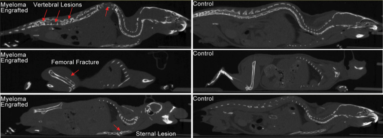
Figure 6. Myeloma-engrafted NSG+hIL6 mice develop skeletal lesions.
MicroCT scans of surviving human IgG+ NSG+hIL6 mice were performed at 52 weeks after injection. Vertebral (top left), femoral (middle left), and sternal (bottom left) lytic lesions in MM1- and MM2-engrafted mice (red arrows) were noted compared with NSG+hIL6 mice not engrafted with MM. Lesions are representative of 14 imaged animals.

