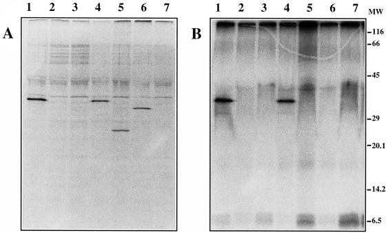
FIG. 8.
Radioimmunoprecipitation analysis of angB-encoded polypeptides in the V. anguillarum cytoplasm. (A) Cultures of V. anguillarum were grown from early log phase to stationary phase in CM9 minimal medium under iron-limiting conditions in the presence of [35S]methionine (50 μCi/ml; ICN Biomedicals Inc.). The cells were collected by centrifugation and processed for immunoprecipitation as described in Materials and Methods. Immunoprecipitated proteins were analyzed by SDS-PAGE and fluorography. Proteins from the following strains were analyzed: lane 1, 531A(pJHC1/pTW99), wild-type strain; lane 2, 531A(pJHC1::Ω/pTW99), phenotype B− G−; lane 3, 531A(pJHC1::K/pTW99), phenotype B− G+; lane 4, 531A(pJHC1::Ω/pTW200), phenotype B− G−/B+ G+; lane 5, 531A(pJHC1::Ω/pTW201), phenotype B− G−/B+ G− (truncated B protein); lane 6, 531A(pJHC1::Ω/pTW200S248L), phenotype B− G−/B+ G−; and lane 7, S531A-1(pTW99), phenotype B− G− (S531A-1 is the plasmidless 531A derivative). (B) Cultures of V. anguillarum were grown from early log phase to stationary phase in a low-phosphate minimal medium under conditions of iron limitation in the presence of 32Pi (500 μCi/ml; ICN Biomedicals Inc.). For this experiment, CM9 medium was modified by replacing phosphate with 100 mM MOPS as a buffer and KH2PO4 was added as a source of inorganic phosphate at a final concentration of 0.3 mM. The labeled cells were processed for immunoprecipitation exactly as described above. Immunoprecipitated 32P-labeled proteins were analyzed by SDS-PAGE and autoradiography. Proteins analyzed were from the same strains as those shown in panel A. MW, molecular mass standards (in kilodaltons).

