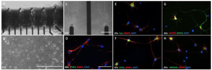
Figure 1. Expression of the molecular targets of AD drugs in hiPSC-derived cortical neurons.
Phase images of hiPSC-derived cortical neurons distributed on patterns aligned with MEA electrodes(A-C), scale bar = 100um..(D-H) Immunocytochemistry of hiPSC-derived cortical neurons utilizing antibodies specific to target proteins relevant for AD drugs revealed the cells expressed all markers evaluated, scale bar = 50um..

