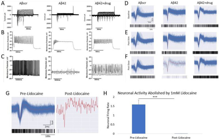
Figure 3. Correlation of the effects of an AD approved drug against Aβ42 oligomers induced neurotoxicity in cortical neurons utilizing two separate functional measurements.
(A-C) Patch clamp electrophysiology recordings indicated a marked reduction in sodium currents in hiPSC-derived cortical neurons following 5 μM Aβ42 oligomer application at 24 hrs post- treatment (A). Additionally, a significant decrease in both induced action potential firing under depolarization (B) and spontaneous firing peak amplitudes (C) was detected in cells treated with Aβ42 relative to cells dosed with amyloid beta scrambled (Aβscr), but the decrease was ameliorated by co-treatment with AD drugs. (D-F) Similar observations for activity in the parallel cortical MEA systems were observed. Following establishment of baseline activity levels (D), the induced LTP activity was maintained at 1hr post dosing (E), and in samples treated with Aβscr (5 μM) no change was observed, a sharp decrease was observed in the samples dosed with Aβ42 oligomers alone within 1hr of treatment and co-treatment of the Aβ42 oligomers and Donepizil rescued the loss of activity (F). (G) Neuronal activity traces and accompanying raster plots illustrating the abolishment of biological activity through blocking of Na+ channels with the addition of 1mM Lidocaine. (H) Graph quantification of G highlighting the abolishment of neuronal signals following lidocaine addition. Statistical analysis was determined via Student’s T-test. *p < 0.05, **p < 0.01, ***p < 0.001.

