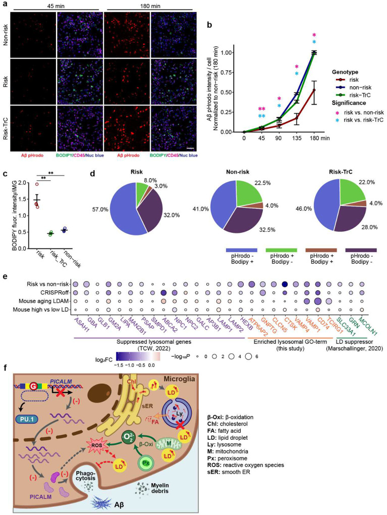
Fig. 6.
LD accumulation impairs iMG phagocytosis and possible mechanism. (a) Representative fluorescence images show time-dependent phagocytosis of Aβ-pHrodo and LD accumulation (BODIPY+) in iMG (CD45+) carrying the LOAD risk allele or non-risk allele of PICALM in the presence or absence of TrC. Scale bar: 100 μm. (b) Reduced Aβ-pHrodo intensity in iMG carrying the LOAD risk allele (vs. non-risk) can be rescued by TrC treatment. (c) TrC treatment rescues the LD accumulation in iMG carrying the LOAD risk allele to a level similar to that in MG carrying the non-risk allele. (d) Pie charts show the proportion of iMG (CD45+) stained positive for Aβ-pHrodo, BODIPY, or both from co-localization analysis of the fluorescence images in (a). Note the Aβ-pHrodo+/BODIPY+ iMG are rare in each type of iMG, and TrC treatment rescues the phagocytosis deficit in iMG carrying the LOAD risk allele by mainly converting BODIPY+ iMG to phagocytic cells without LD (Aβ-pHrodo+/BODIPY−). CD09 line was used, 3 replicate wells each with 1–2 FOV. Student’s t-test (2-tailed, heteroscedastic); * P<0.05, ** P<0.01, ***, P<0.001; error bar, SEM. (e) Dysfunctional lysosome may contribute to LD accumulation in iMG carrying the LOAD risk allele of PICALM. Shown are log2FC of known lysosomal genes and LD suppressor genes in iMG carrying the LOAD risk allele or with PICALM CRISPRoff (vs. non-risk). (f) Mechanistic insight on the link between risk allele of the LOAD GWAS SNP and the reduced PICALM expression, lysosomal dysfunction, LD accumulation, LD peroxidation, cellular ROX level, and phagocytosis deficits in iMG. (−), inhibition; FA: fatty acids; Px: peroxisome; M: mitochondria; sER: smooth ER; Ly: lysosome; Chl: cholesterol.

