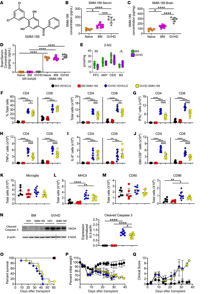
Figure 5. Pharmacological administration of a brain-penetrant CB2R inverse agonist/antagonist reduces inflammation in the CNS during GVHD.
(A) Chemical structure of SMM-189. (B and C) Serum level (B) and brain concentration (C) of SMM-189 in naïve and Balb/c mice transplanted with B6 BM or B6 BM and spleen cells. (D) Ratio of brain to serum SR144528 and SMM-189 concentrations. Results in B–D are from 2 experiments (n = 5–10 mice/group). (E) 2-AG levels in the amygdala, brainstem, cerebellum, and prefrontal cortex 14 days after transplantation. Results are from 3 experiments (n = 14–15 mice/group). (F–N) Balb/c recipients were transplanted with B6 BM alone or B6 BM and spleen cells. Animals were then treated with SMM-189 or vehicle control. (F) The percentage and absolute number of donor CD4+ and CD8+ T cells in the brains of mice 14 days after transplantation. (G–J) The absolute number of CD4+ and CD8+ T cells that produced IFN-γ, TNF-α, IL-6, or GM-CSF. (K) Absolute number of microglial cells. (L and M) Absolute number of MHC class II, CD80, and CD86 expressing microglial cells. Results in panels F–M are from 2 experiments (n = 6–10 mice/group). (N) Representative Western blot images and scatterplots depicting normalized expression of cleaved caspase 3 in the brain from mice treated with either SMM-189 or a vehicle control. Vertical lines on Western blots denote noncontiguous gel lanes. Data are from 2 experiments (n = 4–10 mice/group). (O–Q) Balb/c recipients were transplanted with B6 BM alone (n = 6) or with B6 spleen cells (n = 10) and treated with SMM-189 or vehicle. Overall survival (O), serial weight curves (P), and clinical score (Q) are shown. Results are from 2 experiments (n = 6–10 mice/group). In panels P and Q, BM alone mice only received vehicle. Data are presented as mean ± SD. Statistics were performed using a 1-way ANOVA with Tukey’s test for multiple group comparisons. *P < 0.05, **P < 0.01, ***P < 0.001, ****P < 0.0001. Source data are provided as a Supporting Data Values file.

