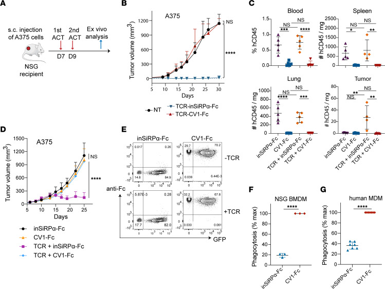
Figure 5. CV1-Fc-engineered T cells are phagocytosed by macrophages in vitro and depleted in vivo.
(A) Schematic of ACT against subcutaneous A375 tumors and ex vivo analysis. (B) A375 tumor growth and control curves following ACT (n = 7). (C) Frequency and number of human CD45+ cells in harvested tissues 5 days after ACT (n ≥4; data are representative of 2 independent studies). (D) A375 tumor growth and control curves following ACT supplemented with coadministration of soluble inSiRPα-Fc and CV1-Fc proteins (n ≥5). (E) Flow cytometry detection of T cell–secreted CV1-Fc binding on T cell surface CD47 by anti–human IgG-Fc Ab staining (data are representative of 6 donors). (F) Phagocytosis of T cells coated with secreted CV1-Fc by NSG BMDMs in vitro (n = 3). (G) Phagocytosis of T cells coated with secreted CV1-Fc by MDMs in vitro (n = 7). Statistical analysis was done by 2-way ANOVA (B and D), 1-way ANOVA (C), or unpaired 2-tailed t test (F and G) with correction for multiple comparisons by post hoc Tukey’s test (B–D). *P < 0.05; **P < 0.01; ***P < 0.001; ****P< 0.0001.

