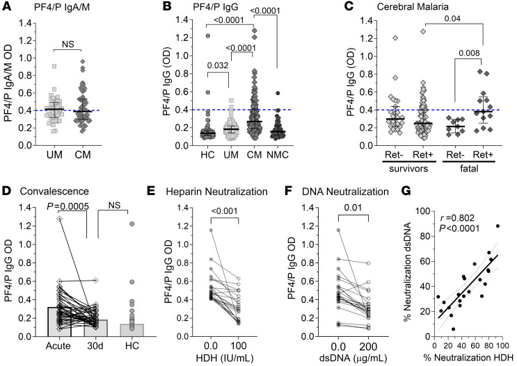
Figure 2. Anti–PF4/P IgG levels are elevated in pediatric CM.
(A) Plasma levels of anti–PF4/P IgM/IgA antibodies in patients with UM (n = 38) versus patients with CM (n = 54). (B) Plasma levels of anti–PF4/P IgG in HCs (n = 56) versus UM (n = 124) versus CM (n = 136) versus NMC (n = 49) patients. (C) Plasma levels of anti–PF4/P IgG in CM survivors (Ret– CM, n = 27; Ret+ CM, n = 87) and patients with fatal CM (Ret– CM, n = 9; Ret+ CM n = 13). (D) Pair-wise comparison of anti–PF4/P IgG in acute versus convalescent plasma from patients with CM 30 days after admission (30d) (n = 39). (E and F) Pairwise analysis of neutralization of anti–PF4/P IgG binding in patient plasma with (E) 100 U/mL HDH (n = 24) or (F) 200 μg/mL dsDNA (n = 24). (G) Pearson’s correlation analysis between neutralization of anti-PF4/P binding by dsDNA versus HDH (n = 22). Shown within the graph is the Pearson’s rho coefficient (r) and the associated P value. In A–C, the assay cutoff threshold of OD above 0.4 is depicted by a dashed blue line. Shown are the median levels ± IQRs. Statistical significance was determined by Mann-Whitney U test (A and D), Kruskal-Wallis test with Dunn’s multiple comparisons (B and C), and a parametric paired t test (E and F).

