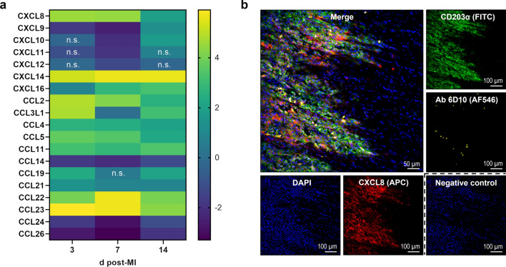
Fig. 10.
Chemokine expression within the infarcted porcine myocardium. a Heat map depicting relative gene expression of CXCL and CCL chemokines determined by bulk RNA sequencing of tissue from the infarct core at the indicated points in time (d = days) post-MI. The color scale shows the log2 fold change vs. tissue from sham hearts (pooled) as calculated in DESeq2 (n = 3–4). n.s. denotes adjusted p > 0.05. b Immunofluorescence staining of CXCL8 and myeloid immune cells in a representative cryosection of myocardial tissue (border zone, 3 days post-MI). CXCL8 (red) is found at the front of infiltrating leukocytes and broadly colocalizes with CD203α+ macrophages (green). Isolated neutrophils (yellow) can be detected by staining with the antibody (Ab) 6D10. The negative control was stained with secondary antibody only. DAPI (4′,6-diamidino-2-phenylindole) was used for nuclear counterstaining (blue)

