Fig. 14.
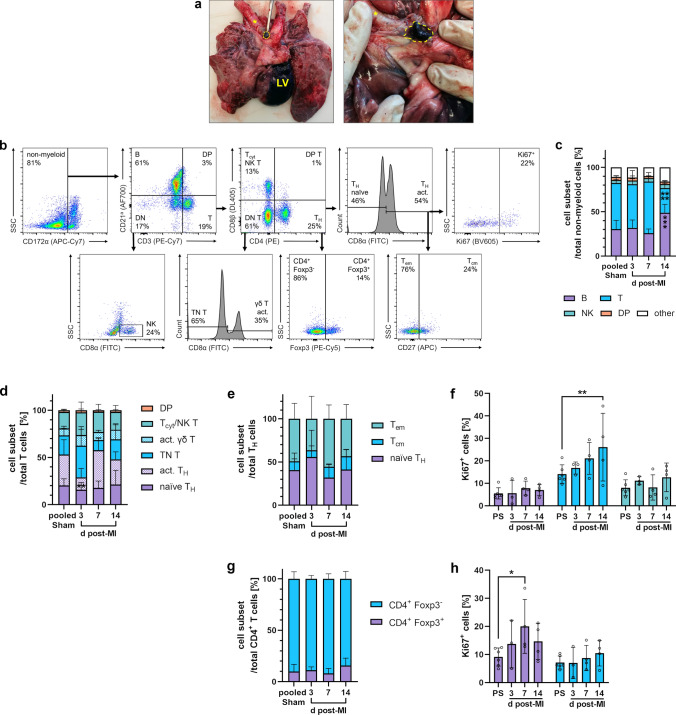
Flow cytometry of lymphocytes in heart-draining lymph nodes of the pig. a Autopsy preparation of the porcine cardiopulmonary system. Mediastinal heart-draining lymph nodes (yellow dashed line) can be identified in close proximity to the trachea (yellow asterisk) after toluidine blue injection into the left-ventricular (LV) myocardium. b Sequential gating strategy for immunophenotyping of lymphocytes. Cells were fixed and permeabilized. All events were pre-gated on viable (Zombie Aqua−) singlets. DN (T): double-negative (T) cells, DP (T): double-positive cells, TN T: triple-negative T cells. c Proportions of lymphocyte subsets at the indicated points in time (d = days) post-MI (n = 3–4) and from sham animals (n = 6). d Proportions of T-cell subsets (n = 3–4 for post-MI, n = 6 for pooled sham). e Proportions of TH subsets (n = 3–4 for post-MI, n = 6 for pooled sham). f Proliferation rate of the individual TH subsets as determined by intracellular Ki67 expression (PS: pooled sham). g Foxp3 intracellular expression on CD4+ T cells (n = 3–4 for post-MI, n = 6 for pooled sham). h Proliferation rate of the individual CD4+ Foxp3± cell subsets. Mean ± SD; *p < 0.05, **p < 0.01, ***p < 0.001, ****p < 0.0001 vs. pooled sham (two-way ANOVA with Dunnett’s multiple comparisons test)

