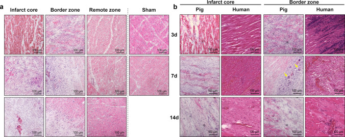
Fig. 2.
Hematoxylin–eosin (H.E.) staining of porcine and human myocardial tissue. a Representative microscopic images from H.E.-stained cryosections of porcine myocardial tissue collected from the different regions at the indicated points in time after MI or sham procedure (d = days). Hypereosinophilia of the necrotic myocardium can be noticed in the infarct core on day 3 post-MI. b Comparison of porcine and human H.E.-stained tissue sections from the infarct core and the border zone. Porcine myocardial tissue was cryoembedded on the indicated days post-MI. Autopsy samples from the hearts of human patients who had died at a comparable point in time after MI were formalin-fixed and paraffin-embedded. Multinucleated giant cells are apparent in the border zone of the pig heart 7 days post-MI (yellow arrows)

