Fig. 3.
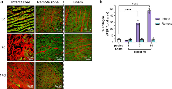
Picrosirius red (PSR) staining of collagen in porcine heart tissue. a Representative fluorescence microscopy images from PSR-stained cryosections collected from the infarct core and the border zone at the indicated points in time after MI or sham procedure (d = days; green autofluorescence: cardiomyocytes, red fluorescence: PSR-positive collagen fibers). b Myocardial collagen content as determined by fluorescence microscopy of PSR-stained tissue cryosections. Mean ± SD; ****p < 0.0001 vs. pooled sham (two-way ANOVA with Dunnett’s multiple comparisons test)

