Fig. 5.
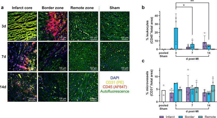
Immunofluorescence staining of leukocytes and microvessels in the porcine myocardium. a Representative immunofluorescence microscopy images from cryosections of myocardial tissue collected from the different regions at the indicated points in time after MI or sham procedure (d = days). Leukocytes were identified by staining for the pan-leukocyte marker CD45 (red). Endothelial cells lining vessels stain positive for CD31 (yellow). DAPI (4′,6-diamidino-2-phenylindole) was used for nuclear counterstaining (blue). Necrotic cardiomyocytes exhibit a distinctive yellow-green autofluorescence clearly different from the less intense green autofluorescence of intact cardiomyocytes. b–c Relative abundance of b leukocytes or c microvessels as determined by immunofluorescence microscopy of stained tissue cryosections. Mean ± SD; *p < 0.05, **p < 0.01 vs. 3d post-MI: border zone (two-way ANOVA with Tukey’s multiple comparisons test)

