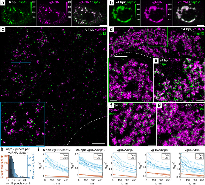
Fig. 3. Association of SARS-CoV-2 replication enzyme with vgRNA clusters.
Representative confocal images of SARS-CoV-2 infected cells display DL colocalization between nsp12, the catalytic subunit of RdRp (green) and vgRNA (magenta) at both 6 hpi (a) and 24 hpi (b). Representative SR images of SARS-CoV−2 infected cells indicate nanoscale association between nsp12 and vgRNA at both 6 hpi (c) and 24 hpi (d). Insets show magnified images of corresponding regions in the colored boxes. Representative SR images of vgRNA with nsp7 (e) or nsp8 (f) in the perinuclear regions of SARS-CoV-2 infected cells indicate association of nsp7 and nsp8 with vgRNA clusters. g Representative SR image of vgRNA with newly synthesized viral RNAs labeled by BrU in a SARS-CoV-2 infected cell indicates localization of newly synthesized viral RNAs within the perinuclear clusters of vgRNA. h Number of nanoscale puncta of nsp12 per vgRNA cluster. Data for the histograms are obtained from 5 cells (6 hpi) and 9 cells (24 hpi). i Bivariate pair-correlation functions for vgRNA and nsp12, nsp7, nsp8 and newly transcribed viral RNA labeled with BrU peak at r = 0 nm indicating association between these target pairs. Scale bars, 10 µm (a, b), 2 µm (c, d), 500 nm (e–g and insets in c and d). Dashed lines in c and d indicate the edge of the cell nucleus.

