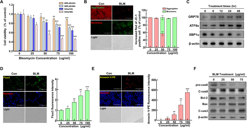
Fig. 1. Impact of bleomycin on ER stress and cell death in cervical cancer cells.
A Viability of WRL68 normal liver and SiHa cervical cancer cells following bleomycin exposure. B Analysis of mitochondrial membrane potential changes using JC-1 staining in response to bleomycin (concentrations: 0, 25, 50, 75, 100 µg/mL), with qualitative (left) and quantitative (right) assessments. C Western blot detection of ER stress markers in cells treated with bleomycin for varying durations (0, 6, 12, 24, 48 hours). D Evaluation of Ca2+ release in cells post-bleomycin treatment, examined through fluorescence microscopy (left) and quantitatively (right) across different dosages. E Assessment of cell apoptosis via Annexin-V staining after bleomycin treatment, visualized through fluorescence microscopy (left) and quantified (right) for each concentration level. F Western blot analysis of apoptosis-related proteins (pro-caspase8, cleaved-caspase8, Bcl2, Bax, cleaved-caspase3) post-treatment with various bleomycin concentrations (0, 25, 50, 75 µg/mL). Significance indicated by *P < 0.05; **P < 0.01; ***P < 0.001.

