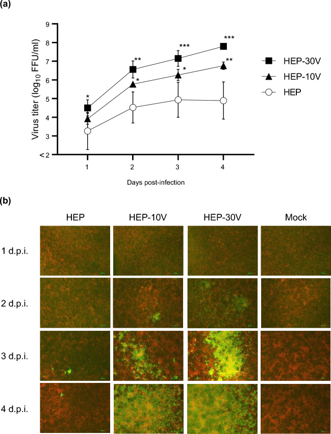
Figure 1.
Viral growth and spread of HEP-Flury (HEP) and Vero-adapted viruses (HEP-10V and -30V) in Vero cells. Vero cells were inoculated with HEP, HEV-10V, or HEV-30V at a multiplicity of infection (M.O.I.) of 0.05. Supernatants were collected once daily through 4 days post infection (d.p.i.). (a) Viral titers were determined using MNA cells. Antigen-positive foci were counted under a fluorescence microscope and quantified as focus forming unit (FFU) per milliliter. Viral titers are plotted as the mean and standard deviation (S.D.) from three independent experiments. Significant differences are indicated (*: p < 0.05, **: p < 0.01, ***: p < 0.001) between HEP and HEP-10V or HEP-30V after application of two-way analysis of variance (ANOVA) followed by Tukey. HEP-10V vs HEP-30V had no significant difference at all time points (p > 0.05). (b) At the indicated time points, virus- and mock-infected cells were fixed with 80% cold acetone. Fixed cells were stained with the fluorescein isothiocyanate (FITC) anti-rabies monoclonal globulin (FUJIREBIO, Tokyo, Japan) and examined under a fluorescence microscope. The stained cells were observed using NIS-Elements D version 5.20.00 imaging software (Nikon, Tokyo, Japan). Cells are stained red with Evans Blue (FUJIFILM Wako Pure Chemical Corporation, Osaka, Japan). Scale bars, 100 μm; magnification, ×40.

