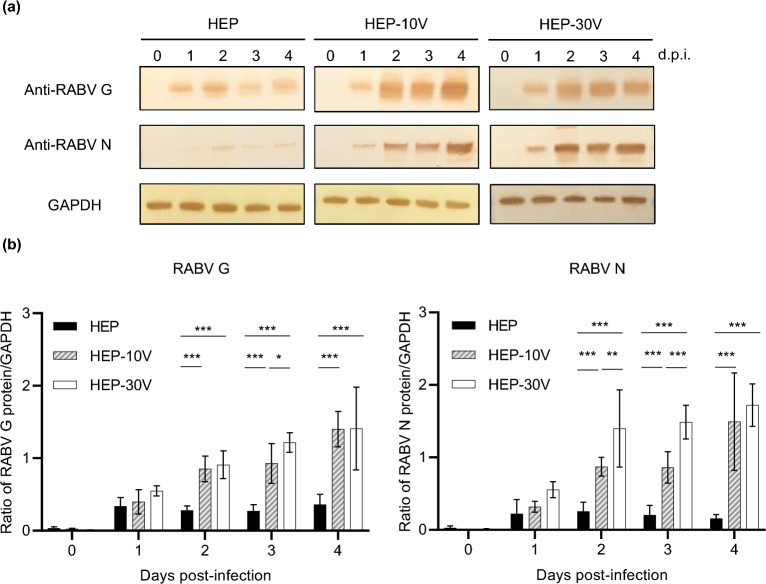
Figure 2.
Comparison of expression of viral proteins among HEP, HEP-10V and HEP-30V in Vero cells. Vero cells were inoculated with each virus at an M.O.I. of 5 and harvested every day. (a) Rabies virus (RABV) glycoprotein (G protein) and nucleoprotein (N proteins) were visualized using polyclonal rabbit anti-serum against each protein. Glyceraldehyde-3-phosphate dehydrogenase (GAPDH), a housekeeping protein, was detected by monoclonal mouse antibody for use as a loading control. (b) Virus-specific bands were quantified using ImageJ software (National Institutes of Health, Bethesda, MD, USA) and levels of RABV G and N proteins were normalized to those of GAPDH. The means and S.D. were calculated from two independent experiments. Significant differences are indicated (*: p < 0.05, **: p < 0.01, ***: p < 0.001) after application of two-way ANOVA followed by Tukey.

