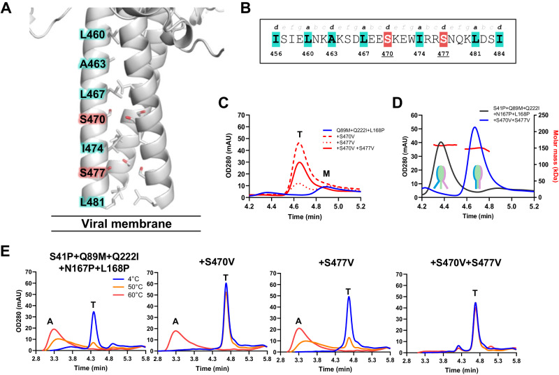
Fig. 2. Stabilization of the RV3 HRB region.
A Cartoon representation of HRB from PDB ID 6MJZ. The heptad register is shown in sticks, with the serines at positions 470 and 477 highlighted in red. B The RV3 F HRB heptad register indicating the suboptimal residues at positions 470 and 477. C Analytical SEC trace of F variants with a minimally stabilized head domain (Q89M, Q222I and L168P) and with or without HRB stabilization in supernatant. The trimer (T) and monomer (M) peaks are indicated. D Analytical SEC-MALS trace of F variants with a stabilized head domain with or without HRB stabilization in supernatant. The molar mass as determined by MALS at peak max of the trimer are indicated. A cartoon to visualize the presumed opening of HRB is shown. E Stability of indicated F variants using analytical SEC of supernatants after 15 min incubation at 4 °C (blue line), 50 °C (orange line), or 60 °C (red line). The trimer (T) and aggregate (A) peaks are indicated.

