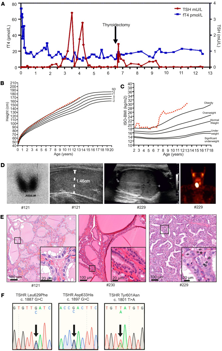
Figure 2. Thyroid function tests, clinical data, imaging results, and genetic data of the patients with de novo germline constitutive active TSHR Leu629Phe mutation and somatic TSHR Asp633His and Tyr601Asn mutations.
(A) Serum thyrotropin (TSH) and free thyroxine (fT4) concentrations of patient #121 from birth to 12 years of age. (B and C) Height-BMI and ISO-BMI curves. (D) Iodine uptake and thyroid ultrasound image at 2 months of age showing high and homogenous radioactivity accumulation in both thyroid lobes and normal-sized thyroid (diameter indicated with white dotted line) in patient #121. Thyroid ultrasound image and single photon emission computed tomography (SPECT) imaging showing exact boundaries of solid thyroid nodule on the right thyroid lobe and large oval accumulation of 99mTc on the right side of thyroid of patient #229. (E) Histological analysis of the H&E-stained thyroid tissue of patients #121, #230, and #229. (F) Representative illustration of Sanger sequencing chromatogram of the patients carrying TSHR Leu629Phe (c.1887G>C, p.l629F), Asp633His (c.1897G>C, p.D633H), and Tyr601Asn (c.1801T>A, p.Y601N) mutations. ISO-BMI, age and sex-adjusted body mass index.

