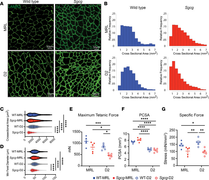
Figure 2. The MRL strain increased myofiber size and maximum tetanic force in Sgcg–/– mice.
TA muscle was analyzed at 20 weeks. (A) Representative immunofluorescence microscopy (IFM) images showed larger myofibers in the TA of the Sgcg-MRL stained for laminin-α2 (LAMA2). Scale bars: 50 μm. (B) Cross-sectional area (CSA) of myofibers was shifted rightward in Sgcg-MRL muscle. (C) CSA was increased in the MRL background (WT-MRL 2544, Sgcg-MRL 2619, WT-D2 2560, and Sgcg-D2 2154 μm2). (D) MRL background increased the minimum Feret diameter in the Sgcg–/– muscle (WT-MRL 49.1, Sgcg-MRL 49.6, WT-D2 49.6, and Sgcg-D2 45.1 μm). (E) Force measurements from male TA muscles showing maximum tetanic force was significantly reduced in the Sgcg-D2 strains compared with all other strains (WT-MRL 1085, Sgcg-MRL 828.5, WT-D2 836.3, and Sgcg-D2 458.7 mN). (F) Physiological CSA (PCSA) was smaller in D2 compared with MRL muscle for both Sgcg–/– and WT (WT-MRL 7.44, Sgcg-MRL 8.14, WT-D2 5.1, and Sgcg-D2 4.85 mm2). (G) Specific force was reduced for Sgcg–/– compared with WT in the D2 strain but not the MRL strain (WT-MRL 145, Sgcg-MRL 102.3, WT-D2 163.5, and Sgcg-D2 96.0 mN/mm2). Data are presented as mean ± SD. *P < 0.05; **P < 0.01; ***P < 0.001; ****P < 0.0001 by 2-way ANOVA with Tukey’s multiple-comparison test.

