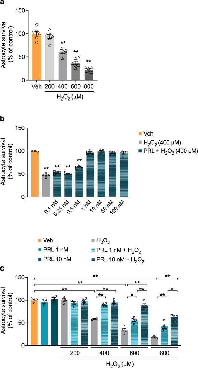
Fig. 1.

Effect of PRL on H2O2 toxicity in rat astrocytes. a Rat cortical astrocytes were incubated for 3 h in the absence or presence of increasing concentrations (200–800 μM) of hydrogen peroxide (H2O2). Cell viability was quantified by MTT assay, and the results were normalized to the control with vehicle. Data are means ± SEM of six independent primary cultures (n = 6) carried out in triplicate. b Rat cortical astrocytes were pre-incubated for 24 h in the absence or presence of increasing concentrations (1–100 nM) of prolactin (PRL), then 400 μM H2O2 or vehicle was added and incubated for 3 h. Cell viability was quantified by MTT assay, and the results were normalized to the control with vehicle. Data are means ± SEM of five independent primary cultures (n = 5) carried out in triplicate. c Rat cortical astrocytes were pre-incubated for 24 h in the absence or presence of 1 or 10 nM PRL, then 400 μM H2O2 or vehicle was added and incubated for 3 h. Cell viability was quantified by MTT assay, and the results were normalized to the control with vehicle. Data are means ± SEM of four independent primary cultures (n = 4) carried out in triplicate. One-way ANOVA followed by Tukey’s test; *p < 0.05, **p < 0.001 versus vehicle or indicated group
