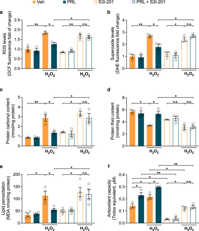
Fig. 3.
Effect of PRL on H2O2-induced ROS production and oxidative damage in rat astrocytes. Rat cortical astrocytes were pre-incubated in the absence or presence of 10 nM prolactin (PRL),100 μM STAT3 inhibitor (S3I-201) or both for 24 h, then 400 μM hydrogen peroxide (H2O2) or vehicle (Veh) was added and incubated for 3 h. a Generation of reactive oxygen species (ROS) was quantified using 2′,7′-dichlorodihydrofluorescein diacetate (DCF-DA). Values are expressed as DCF fluorescence after 30 min of incubation. Data are means ± SEM of three independent experiments (n = 3) carried out in triplicate. b Generation of superoxide anion was measured using dihydroethidium (DHE). Values are expressed as DHE fluorescence after 30 min of incubation. Data are means ± SEM of three independent experiments (n = 3) carried out in triplicate. c Protein oxidation was estimated by measuring the protein carbonyl levels with the DNPH colorimetric assay. The concentration of the protein carbonyls was adjusted to the total protein concentration. Data are means ± SEM of three independent experiments (n = 3) carried out in duplicate. d Total sulfhydryl groups were measured by the reaction of free thiols in native proteins with DTNB. The concentration of the free thiols was adjusted to the total protein concentration. Data are means ± SEM of three independent experiments (n = 3) carried out in duplicate. e Lipid peroxidation was determined as the increase in malondialdehyde (MDA), a thiobarbituric acid reactive substance (TBARS). The concentration of the MDA was adjusted to the total protein concentration. Data are means ± SEM of four independent experiments (n = 4). f Antioxidant capacity was detected by ABTS assay. Data are means ± SEM of three independent experiments (n = 3) carried out in duplicate. One-way ANOVA followed by Tukey’s test; *p < 0.05, **p < 0.001 versus indicated group; n.s. no significant difference

