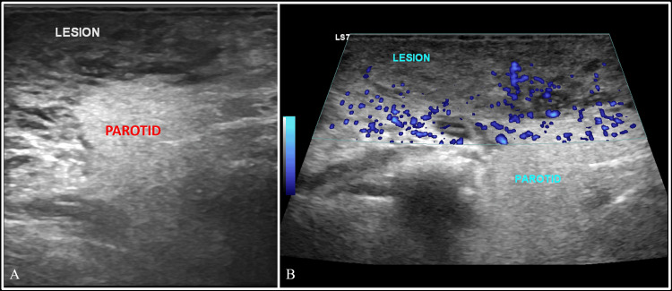
Figure 1. Greyscale ultrasound of the neck.
Greyscale ultrasound (A) and power doppler (B) images of the neck at the infraauricular level on the right side, showing a large heteroechoic mass superficial to the parotid gland, showing few areas of vascularity within it. Note the difference in the echotexture between the soft tissue mass and the parotid.

