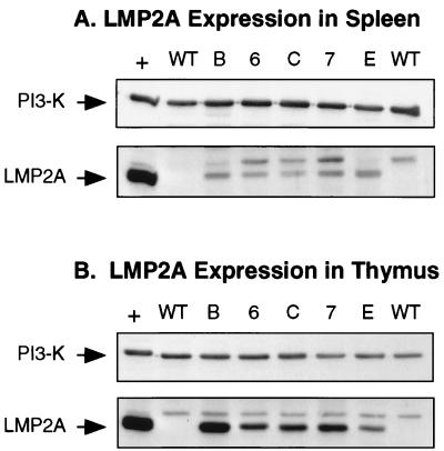
FIG. 1.
Immunoblot detection of LMP2A expression in EμLMP2A spleen (A) and thymus (B) tissues. Whole spleen and thymus organs from EμLMP2A animals were excised and dissociated between frosted slides. Membranes were cut in half, allowing independent analysis of the same sample with LMP2A (45 kDa) and PI3 kinase (85-kDa subunit) antibodies. The bottom portion of each membrane was probed with rat 14B7 anti-LMP2A monoclonal antibody. The top portion of each blot was probed with rabbit anti-p85 primary antibody. Cell lysates from LCL10 cell equivalents were used as a positive control (+) for LMP2A expression. The LCL10 positive control lane contains 0.5 × 10⁶ cell equivalents; all EμLMP2A lanes contain 2.5 × 10⁶ cell equivalents. Wild-type animals are as indicated (WT). EμLMP2A genotypes are indicated by single alphanumeric abbreviations.

