



Abstract
AIM
To compare high or low concentration of hyaluronic acid eye drops (HY) for dry eye syndromes (DES).
METHODS
Randomized controlled trials (RCTs) comparing various concentrations of HY were searched in PubMed, Embase, Web of Science, Cochrane, SinoMed, CNKI, Wanfang Database, CQVIP, and Chinese journals databases between inception and July 2023. Pooled standardized mean differences (SMD) or weighted mean difference (WMD) with 95% confidence intervals (CI) from RCTs evaluating Schirmer's I test (SIT), corneal fluorescein staining score (CFS), tear breakup time (TBUT), DES score (DESS), and Ocular Surface Disease Index (OSDI) were calculated. Sensitivity analysis, Egger's test and Meta-regression analysis were performed for all indicators.
RESULTS
We conducted a Meta-analysis of 10 RCTs that met the inclusion criteria, involving 1796 cases. High-concentrations group significantly improved the outcome of CFS according to random effects modelling (SMD, -3.37; 95%CI, -5.25 to -1.48; P=0.0005). The rest of the results were not statistically significant, including indicators such as SIT, TBUT, DESS and OSDI.
CONCLUSION
For dry eyes with positive corneal staining, a high concentration of HY is recommended, whereas in other cases, a high concentration of HY does not offer a more pronounced advantage over a low concentration of HY in the treatment of dry eyes.
Keywords: dry eye, hyaluronic acid, concentration, Meta-analysis
INTRODUCTION
Dry eye syndrome (DES) is a complex ocular surface disease that involves multiple factors. According to the International Dry Eye Workshop, dry eye is characterized by an imbalance in the tear film and ocular symptoms. However, due to reduced corneal sensitivity, such patients may also have tear abnormalities without symptoms. The various factors contributing to the development of dry eye often overlap and interact, including tear-film instability, hyperosmolarity, ocular surface inflammation and damage, and neurosensory abnormalities[1]. Additionally, dry eyes can harm visual function and quality of life and impose a significant economic burden[2]–[3].
Various factors can contribute to the development of dry eye. Certain diseases, such as diabetes, can lead to damage in the microvasculature of the lacrimal gland, autonomic neuropathy, and diabetic sensory neuropathy of the cornea, all of which can impact the quality and quantity of tears[4]. Additionally, chronic graft-versus-host disease accompanied by conjunctival inflammation and fibrosis[5], as well as autoimmune diseases like rheumatoid arthritis, systemic lupus erythematosus, and Sjögren's syndrome, can also contribute to dry eye[6]. Furthermore, impairment of the neural feedback loop responsible for regulating tear secretion can worsen the symptoms of ocular surface disease[7]. Obstructive meibomian-gland dysfunction can alter the lipid composition of tears[8], while conjunctival achalasia and eyelid laxity (floppy eyelid syndrome) may also contribute to the development of dry eye[9]. Certain medications, such as furosemide, propranolol, candesartan, cetirizine, and ranitidine, have been known to trigger dry eye[10]. Some topical ocular medications contain preservatives, such as benzalkonium chloride, that can cause or worsen dry eye symptoms and signs[11]. It is imperative to remain cognizant of other potential instigators for dry eyes, such as ocular-surface inflammation caused by ocular disease, infection, or immune-mediated conditions, as well as environmental exposures such as wind and airborne particulates[12]. Moreover, sex hormones can exert an impact on the surface of the eye by modifying goblet-cell density and the production and quality of tears[13]. Any ailment or circumstance that diminishes the blink rate can intensify the likelihood of dry eye by inducing tear evaporation, such as Parkinson's disease or prolonged screen viewing[14]–[16].
To effectively manage DES, several therapeutic strategies can be employed. These strategies include reducing inflammation, modifying one's diet and lifestyle choices, and treating any associated eyelid conditions. In addition, the use of artificial tear formulations of different viscosities and compositions[17], topical lubricants in the form of gels or ointments, diquafosol tetrasodium ophthalmic solution, and autologous serums can all improve DES. Among these formulations, sodium hyaluronate is a frequently utilized artificial tear that can increase retention time and improve ocular surface hydration and lubrication, ameliorating DES. Clinical trials have demonstrated the efficacy of sodium hyaluronate eye drops as a viable treatment option for DES[18]. With the earliest clinical trials dating back to 1986[19], hyaluronic acid eye drops (HY) can be considered an “old hero” in the fight against dry eye disease. Previous Meta-analyses have demonstrated that preparations containing HY improve Schirmer's I test (SIT) more than non-HY preparations and less improvement in tear breakup time (TBUT)[20]. However, no literature compares explicitly the effects of HY concentration.
Currently, the concentration of HY ranges from 0.1% to 0.4%, and most clinical trials have shown that the higher the concentration of HY, the more effective it is. This Meta-analysis aimed to compare the efficacy of high-concentration HY versus low-concentration HY preparations in the treatment of DES and to determine whether there is an advantage to increasing the concentration from an evidence-based perspective. No evidence-based research articles were found at the time of closing this article. In addition, this paper was registered with PROSPERO under registration number CRD42023453696.
MATERIALS AND METHODS
Search Strategy
Our research team has conducted a comprehensive search of English and Chinese language databases, utilizing a variety of search terms on “Hyaluronic Acid” and “Dry Eye Syndromes”. We have thoroughly examined PubMed, Embase, Web of Science, and the Cochrane Database, as well as SinoMed, CNKI, Wanfang Database, CQVIP, and Chinese Med journals. Our search has encompassed all pertinent publications from inception to July 2023 without imposing any language restrictions.
Selection Criteria
Our review adhered to the PRISMA guidelines and utilized a predetermined protocol. Our inclusion criteria consisted solely of randomized controlled trials (RCTs) conducted on human subjects, focusing on comparing the efficacy of various concentrations of hyaluronic acid. In cases where additional interventions were administered (e.g., tobramycin dexamethasone eye drops), all groups had to be identical. Two authors (Ouyang XW, Fang S) independently confirmed the eligibility of the studies and collated the data from the qualifying studies. Data extracted by Ouyang XW and Fang S, both sides check each other out, and the senior author (Yi YM) is responsible for resolving discrepancies. This article does not discuss any ethical concerns.
Statistical Analysis
The Meta-analyses were performed using Revman 5.4 and Stata17 Software. Standardised mean difference (SMD) or weighted mean difference (WMD) was used for continuous variables, and 95% confidence intervals (CI) were used to calculate pooled estimates. A statistically significant P-value was considered to be less than 0.05. Heterogeneity between trials was measured using I2 values, with I2 values greater than 50% indicating significant heterogeneity. The random effects model analyzed all the results, and sensitivity analysis and Meta-regression methods were used for the included results. Finally, Egger's test was used to determine publication bias, and trim and fill analysis were employed if publication bias was detected.
RESULTS
Literature Search Results
The initial literature search identified a total of 6039 articles. Following the removal of 2502 duplicates, 3527 articles were excluded based on the inclusion criteria. The remaining ten studies were retrieved successfully and were incorporated in the Meta-analysis. Of these, 8 RCTs reported TBUT results[21]–[28]. It is important to note that since TBUT was the sum of three times in the study of Calonge et al[21], we did not include this literature, and since mild and severe phenotypes were compared in the study of Zheng and Zhao[28], we collected two sets of data from this study. Three RCTs[21],[24],[28] reported Ocular Surface Disease Index (OSDI) results, and two sets of data were reported by type in the Zheng and Zhao's study[28]. Four RCTs[21],[23],[25]–[26] reported SIT results, while 4 RCTs[22]–[23],[26],[29] reported DES score (DESS) results. Five RCTs[22]–[24],[26],[30] reported corneal fluorescein staining score (CFS) results. Figure 1 summarizes the study selection process.
Figure 1. Selection process for the study.

Characteristics of Studies
This review encompasses ten studies conducted between 2014 and 2023, which investigate the effects of various interventions on individuals with DES. The study population is diverse, including postoperative cataract surgery, laser in situ keratomileus surgery (LASIK), and non-surgical cases. Study participants exhibit varying degrees of severity of DES and are followed up for a period ranging from 4 to 12wk. The concentration of HY administered across these studies ranges from 0.1% to 0.3%. In addition, 5 of the articles include other interventions, such as antiphlogosis eye drops, and these interventions are consistent across all groups. Table 1 provides a summary of crucial study characteristics.
Table 1. Summary of crucial study characteristics.
| No. | Study | Location | Period | Population | Age, (mean+SD, y) | Follow up duration | Severity of DES of subject | Concentration of HY | Other interventionsb | SIT | TBUT | DESS | CFS | OSDI |
| 1 | Calonge et al[21] | Spain | 2023 | Non-surgical | 55.7±12.4 | 5-12wk | Moderate to severe | 0.18% vs 0.3%a | None | Yes | Yesc | No | No | Yes |
| 2 | Hong and Chen[22] | China | 2014-2015 | Postoperative cataract surgery | 51-78 (range) | 4wk | Unspecified | 0.1% vs 0.3% | Tobradex+pranopulin | No | Yes | Yes | Yes | No |
| 3 | Jin and Dang[23] | China | 2015-2016 | Postoperative cataract surgery | 68.63±5.93 | 1mo | Unspecified | 0.1% vs 0.3% | Tobradex+pranopulin | Yes | Yes | Yes | Yes | No |
| 4 | Jun et al[24] | Korean | 2022 | Non-surgical | 46.1±14.3 | 4-8wk | Moderate to severe | 0.15% vs 0.3% | None | No | Yes | No | Yes | Yes |
| 5 | Ntonti et al[25] | Greece | 2017-2018 | Postoperative cataract surgery | 72.4±8.7 | 4-6wk | Non-DES | 0.1% vs 0.3% | Tobradex | Yes | Yes | No | No | No |
| 6 | Song[26] | China | 2016-2017 | Postoperative cataract surgery | 66.12±1.14 | 1mo | Unspecified | 0.1% vs 0.3% | Tobradex+pranopulin | Yes | Yes | Yes | Yes | No |
| 7 | Yin and Kong[27] | China | 2020-2021 | Post-LASIK | 25.11±1.21 | 1mo | Unspecified | 0.1% vs 0.3% | None | No | Yes | No | No | No |
| 8 | Zheng and Zhao[28] | China | 2016-2017 | Post-LASIK | 24.1±5.0 | 1-3mo | Mild and severe | 0.1% vs 0.3% | None | No | Yesd | No | No | Yesd |
| 9 | Jing[29] | China | 2018 | Postoperative cataract surgery | 64.57±4.35 | 1mo | Unspecified | 0.1% vs 0.3% | Tobradex+pranopulin | No | No | Yes | No | No |
| 10 | Yang[30] | China | 2017 | Postoperative cataract surgery | 56±4.6 | 1mo | Unspecified | 0.1% vs 0.3% | None | No | No | No | Yes | No |
HY: Hyaluronic acid eye drops; SIT: Schirmer's I test; TBUT: Tear break-up time; DESS: Dry eye symptom score; CFS: Corneal fluorescein staining score; OSDI: Ocular Surface Disease Index; LASIK: Laser in situ keratomileus surgery; DES: Dry eye syndrome; Tobradex: Tobramycin dexamethasone eye drops. aHyaluronic acid 0.30% ocular gel; bDrugs were the same in each group in the single study; cSum of 3 measurements; dTwo groups of cases were collected.
Risk of Bias Assessment
Figure 2 shows the results of the risk of bias assessment for the 10 RCTs for which Meta-analysis was subsequently performed. Among the 10 studies, 6 RCTs reported appropriate random sequence generation, and 7 RCTs did not detail allocation concealment or blinding methods. No biases related to attrition or other aspects were detected in any of the studies. One RCT found a high risk of selection bias, and another identified a high risk of reporting bias. All studies featured in the Meta-analysis underwent assessment of potential bias employing the Cochrane Risk of Bias tool[31].
Figure 2. The results of the risk of bias assessment.

Meta-Analysis Results
Baseline data
Upon analysis of the baseline data of age, SIT, TBUT, DESS, CFS, and OSDI, it observes that none of the outcomes were statistically significant. In addition, in the analysis of heterogeneity, the value of age was 53%, and the rest of the results were less than 50%, with little overall heterogeneity (Figure 3).
Figure 3. Forest plot results for each baseline data.

Schirmer's I Test
Based on the analysis conducted through random-effects modeling, there was no significant improvement in SIT outcome within the high-concentration group compared to those using low-concentration preparations. The WMD was calculated to be 0.57, with a 95%CI of -0.15 to 1.29, and the data did not achieve statistical significance (P=0.12). There was significant overall heterogeneity with an I2 value of 80%. Moreover, Egger's test did not indicate publication bias (P=0.795; Figure 4).
Figure 4. Forest plot and Egger's test of Schirmer's I test.

Tear Breakup Time
The analysis shows no substantial variation in the TBUT outcome between the high-concentration group and the low-concentration group. The random-effects modeling showed the WMD was calculated to be 0.98, with a 95%CI of -1.09 to 3.04, and the results did not reach statistical significance (P=0.35). There was significant overall heterogeneity with an I2 value of 98%. Egger's test demonstrated significant asymmetry (P=0.045), while the trim and fill analysis did not have any impact on the results, indicating stable TBUT outcomes (Figure 5).
Figure 5. Forest plot and Egger's test, Trim and Fill analysis of tear breakup time.

Dry Eye Symptoms Score
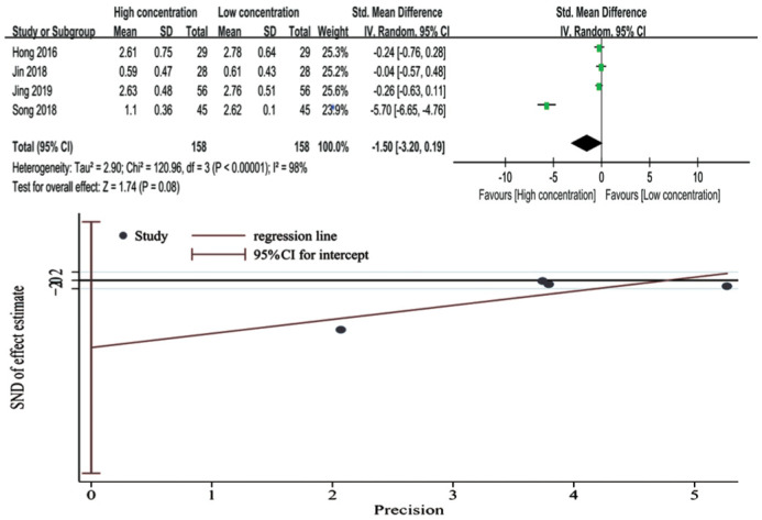
The results of the analysis using random-effects modeling indicate that the high-concentration group did not demonstrate any significant improvement in DESS outcome when compared to the low-concentration preparations. The SMD was calculated to be -1.50 with a 95%CI ranging from -3.20 to 0.19, and the results did not reach statistical significance (P=0.08), the overall heterogeneity was found to be significant (I2=98%). Egger's regression test analysis revealed no evidence of publication bias (P=0.148; Figure 6).
Figure 6. Forest plot and Egger's test of dry eye symptom score.
Corneal Fluorescein Staining Score
Random-effects modeling demonstrated that individuals in the high-concentration group experienced a notable improvement in CFS outcome compared to those utilizing low-concentration preparations. The SMD was determined to be -3.37 with a 95%CI of -5.52 to -1.48, and the results were statistically significant (P=0.0005), the significant overall heterogeneity with an I2 value of 98%. Egger's regression test showed significant asymmetry with statistical significance (P=0.000). However, after implementing the trim and fill method, it was determined that the combined results did not change direction. The P value before and after cutting and filling was 0, indicating robust results (Figure 7).
Figure 7. Forest plot and Egger's test, Trim and Fill analysis of corneal fluorescein staining score.

Ocular Surface Disease Index
The analysis results reveal that there is no significant variation in OSDI outcome between high-concentration and low-concentration groups as per the random-effects model. The SMD stands at -0.24 with a 95%CI of -0.75 to 0.27 and the results did not attain statistical significance (P=0.36). The overall heterogeneity was significant, with a value of 64%. Moreover, Egger's regression test demonstrated no publication bias (P=0.857). According to region, Meta-regression analysis did not identify the source of heterogeneity (Figure 8).
Figure 8. Forest plot and Egger's test of ocular surface disease index.

Sensitivity Analysis and Meta-regression
We performed sensitivity analyses to analyze the heterogeneity factors. The results were all between the upper and lower limits of the CI, and the conclusions were relatively stable. Only the SIT result showed a significant rightward shift in the CI (Figure 9). Further, we performed Meta-regression and did not find the source of heterogeneity (Figure 10).
Figure 9. Sensitivity analysis of each result.

SIT: Schirmer's I test; TBUT: Tear break-up time; DESS: Dry eye symptom score; CFS: Corneal fluorescein staining score; OSDI: Ocular Surface Disease Index.
Figure 10. Meta-regression of each result.

DISCUSSION
It is widely recognized that DES arises from reduced tear secretion or increased tear evaporation, leading to conditions such as aqueous-deficient dry eye and evaporative dry eye[32]–[33]. HY is increasingly being used in patients with all types of dry eye due to its excellent hydrating and lubricating capabilities. A prior Meta-analysis demonstrated a noteworthy enhancement in the SIT test score subsequent to using HY eye drops when compared to non-HY treatment[20]. However, no statistically significant results were observed, including for TBUT and OSDI[34], and there were no comparison of the impact of changes in concentration on dry eye symptoms. This study mainly compared the effect of high concentration of HY (0.3%) and low concentration of HY (0.1%, 0.15%, 0.18%) in treating dry eye patients, including SIT, TBUT, CFS, DESS, and OSDI results, whereas none of the previous Meta-analyses compared the effect of high and low concentrations of HY eye drops on dry eye.
Generally, low-concentration of HY is suggested for mild DES, and high-concentration of HY is advised for severe DES. This article provides some evidence of the superiority of high-concentration HY in the treatment of DES. The CFS significantly decreased in the high-concentration group compared to the low-concentration group (SMD -3.37; 95%CI -5.25, -1.48) with high heterogeneity (I2=98%, P<0.00001). Similarly, relevant animal studies demonstrating significantly lower CFS in the higher concentration HY group compared to the lower concentration groups[35]. However, none of the other results we analyzed were statistically significant, but through forest plots of DESS and OSDI we found that high concentration group is favoured. We initially thought that a higher concentration of HY would be more beneficial. Although some academics have argued that characterizing low concentration of HY as sufficient for mild cases of dry eyes, while higher concentration are better suited to severe instances, this argument is inaccurate[36]–[37].
The SIT, which quantifies tear production, is a prevalent diagnostic technique in DES[38]. The SIT did not yield any statistically significant difference between the high and low concentration groups (WMD 0.57; 95%CI: 0.15, 1.29) as well as high heterogeneity (I2=80%, P=0.002). We identified the study conducted by Jin and Dang[23], which seemed to be responsible for the observed heterogeneity. By excluding this study, the heterogeneity was reduced to 0 (I2=0) and significant changes were observed (P<0.00001). However, the Meta-regression analysis did not reveal the source of heterogeneity. For one thing, this may have to do with the limitations of testing. The absence of standardised placement of paper strips, along with uneven absorption upon tearing, the imprecise correlation between liquid absorption and humidification length of the strip, and the deficiency of standardised assessment methods all contribute to low reproducibility, sensitivity, and specificity of SIT. This may lead to inaccurate results[39]. On the other hand, clinicians often rely on the SIT to determine whether the eye is aqueous-deficient dry eye. Since none of the studies we included differentiated between aqueous-deficient dry eye and evaporative dry eye and HY does not belong to the class of medications that promote tear secretion, it is not unlikely that the SIT is not statistically significant. Although some researchers have argued that the SIT is not sensitive enough to be useful as a tool for patients with non-severe DES[40]. Nevertheless, the SIT continues to be used as a standard diagnostic tool for DES monitoring.
Like the SIT, TBUT results were also statistically insignificant (P=0.35) and demonstrated high heterogeneity (I2=98%), but sensitivity analysis showed that the results were stable. TBUT mainly evaluates the tear film in cases of evaporative DES[41]. A previous Meta-analysis demonstrated a lesser improvement in TBUT following the use of a hyaluronan-based treatment[42]. From the results of the current study, the increase in HY concentration also did not improve TBUT. It may be due to the instability of the tear film itself. False tear film rupture may occur as a result of lipid layer diffusion and absorption into the mucus-water interface or due to the rupture of the least strong region of the mucus layer[43]. Simultaneously, the test has no standardized procedure for the application of fluorescein, which can lead to inaccurate results. Although it has been labeled inaccurate and non-reproducible[44], it is still recommended to be routinely assessed in clinical practice by measuring TBUT[45].
Both DESS and OSDI reflect the eye's subjective perception, and the scores are largely limited by the patient's tolerance and corneal sensitivity. It is established that such subjective test yields more dependable and replicable outcomes than alternative objective measures[46]. Although neither metric was statistically significant, the forest plot favoured the higher concentration group, so we think it still proves the advantage of the higher concentration group. However, corneal nerve damage resulting from prolonged DES is a well-established phenomenon. This can lead to reduced corneal sensitivity, which may obscure symptoms of discomfort within the eye[47]. At the same time, the selection of rating scales was not standardized, and the degree of dry eye in the study population was different. All of these factors affect the accuracy of the final scoring results. Further, Egger's regression test revealed no evidence of publication bias, and Meta-regression analyses failed to identify the origin of heterogeneity, thus indicating relatively dependable outcomes.
Combining the above results, this is probably related to the physicochemical properties of HY. Inflammation has been identified as the pathogenic mechanism of dry eye disease[48]. High molecular weight hyaluronic acid is an anti-inflammatory mediator, while low molecular weight hyaluronic acid is a pro-inflammatory mediator[49]–[51]. Therefore, HY plays an important role in patients with moderate or severe dry eye and superficial keratitis, especially in patients with dry eye accompanied by inflammation[52]. The impact of HY on DES treatment is attributable to diverse mechanisms of action, including stabilizing the ocular surface barrier and tear film, reducing mechanical damage of the cornea[53], increasing cell adhesion and motility[54] and promoting cellular migration[55], and reducing tear evaporation[56]. Hence the choice of treating dry eye with HY preparations.
However, raising the concentration of HY alone may not provide the anticipated benefits. Due to variations in molecular weight and polydispersity index among the HY formulations utilized, as well as other physico-chemical characteristics, which can have a notable influence on the overall viscosity and clinical applications of HY[57]. HY solutions are commonly prescribed as the major treatment for moderate to severe DES in clinical practice, due to their anti-inflammatory and immunomodulatory properties[58], but the effect varies from person to person. As a result, drug companies attempt to improve the efficacy of HY by increasing the concentration and improving the ratio, and of course this behaviour is beneficial.
The study's primary limitation concerns the heterogeneity of the included studies. Differences in research design between studies (random assignment method and blind method), characteristics of subjects (differences in age, follow-up period, region, requires surgery, other intervention) in particular severity of DES, and whether hyaluronic acid contains preservatives are not clearly stated in the vast majority of literature. The original study also did not specify when the test was performed (how long after the eye drops were administered). These factors could have influenced the outcomes of this research. Based on it. First, Table 1 provides a detailed overview of the critical characteristics of each study to enhance the contextualization of our findings. Second, we tried to find points of difference (region, other interventions) in the literature for each outcome, looking for sources of heterogeneity through Meta-regression. Finally, we chose a follow-up period of 4-5wk for all included studies to minimize the effect of this factor. Because the one with a 5-week follow-up was a high-quality study, we did not exclude it.
This review analyzed ten clinical trials that compared the effectiveness of various concentrations of HY. The Meta-analysis demonstrated that high concentrations of HY were more effective in improving CFS than low concentrations. However, they were found to be ineffective in improving other indicators, particularly SIT and TBUT. Although the high concentration group showed some benefits in DESS and OSDI, which may enhance the subjective experience of patients, it was not statistically significant. Therefore, it is advisable to use higher concentrations of HY for the treatment of dry eye with corneal staining. The main limitation of this study is the inter-study heterogeneity, which suggests that a prominent human RCT with a standardized protocol is needed to properly assess the relative efficacy of high-concentration versus low-concentration artificial tear preparations.
Footnotes
Conflicts of Interest: Ouyang XW, None; Fang S, None; Yi YM, None; Zou SP, None; Hu QY, None; Huang ZX, None; Li QX, None; Luo JY, None.
REFERENCES
- 1.Sheppard J, Shen Lee B, Periman LM. Dry eye disease: identification and therapeutic strategies for primary care clinicians and clinical specialists. Ann Med. 2023;55(1):241–252. doi: 10.1080/07853890.2022.2157477. [DOI] [PMC free article] [PubMed] [Google Scholar]
- 2.Okumura Y, Inomata T, Iwata N, Sung J, Fujimoto K, Fujio K, Midorikawa-Inomata A, Miura M, Akasaki Y, Murakami A. A review of dry eye questionnaires: measuring patient-reported outcomes and health-related quality of life. Diagnostics. 2020;10(8):559. doi: 10.3390/diagnostics10080559. [DOI] [PMC free article] [PubMed] [Google Scholar]
- 3.Boboridis KG, Messmer EM, Benítez-Del-Castillo J, Meunier J, Sloesen B, O'Brien P, Quadrado MJ, Rolando M, Labetoulle M. Patient-reported burden and overall impact of dry eye disease across eight European countries: a cross-sectional web-based survey. BMJ Open. 2023;13(3):e067007. doi: 10.1136/bmjopen-2022-067007. [DOI] [PMC free article] [PubMed] [Google Scholar]
- 4.Zhou QJ, Yang LL, Wang Q, Li Y, Wei C, Xie LX. Mechanistic investigations of diabetic ocular surface diseases. Front Endocrinol. 2022;13:1079541. doi: 10.3389/fendo.2022.1079541. [DOI] [PMC free article] [PubMed] [Google Scholar]
- 5.Carreno-Galeano JT, Dohlman TH, Kim S, Yin J, Dana R. A review of ocular graft-versus-host disease: pathophysiology, clinical presentation and management. Ocul Immunol Inflamm. 2021;29(6):1190–1199. doi: 10.1080/09273948.2021.1939390. [DOI] [PubMed] [Google Scholar]
- 6.Shah R, Amador C, Tormanen K, Ghiam S, Saghizadeh M, Arumugaswami V, Kumar A, Kramerov AA, Ljubimov AV. Systemic diseases and the cornea. Exp Eye Res. 2021;204:108455. doi: 10.1016/j.exer.2021.108455. [DOI] [PMC free article] [PubMed] [Google Scholar]
- 7.Hat K, Kaštelan S, Planinić A, Muller D, Ježek D. Pathohistological features of the aging human lacrimal gland. Croat Med J. 2023;64(5):307–319. doi: 10.3325/cmj.2023.64.307. [DOI] [PMC free article] [PubMed] [Google Scholar]
- 8.Sheppard JD, Nichols KK. Dry eye disease associated with meibomian gland dysfunction: focus on tear film characteristics and the therapeutic landscape. Ophthalmol Ther. 2023;12(3):1397–1418. doi: 10.1007/s40123-023-00669-1. [DOI] [PMC free article] [PubMed] [Google Scholar]
- 9.Salinas R, Puig M, Fry CL, Johnson DA, Kheirkhah A. Floppy eyelid syndrome: a comprehensive review. Ocul Surf. 2020;18(1):31–39. doi: 10.1016/j.jtos.2019.10.002. [DOI] [PubMed] [Google Scholar]
- 10.Askeroglu U, Alleyne B, Guyuron B. Pharmaceutical and herbal products that may contribute to dry eyes. Plast Reconstr Surg. 2013;131(1):159–167. doi: 10.1097/PRS.0b013e318272a00e. [DOI] [PubMed] [Google Scholar]
- 11.Goldstein MH, Silva FQ, Blender N, Tran T, Vantipalli S. Ocular benzalkonium chloride exposure: problems and solutions. Eye (Lond) 2022;36(2):361–368. doi: 10.1038/s41433-021-01668-x. [DOI] [PMC free article] [PubMed] [Google Scholar]
- 12.Perez VL, Stern ME, Pflugfelder SC. Inflammatory basis for dry eye disease flares. Exp Eye Res. 2020;201:108294. doi: 10.1016/j.exer.2020.108294. [DOI] [PMC free article] [PubMed] [Google Scholar]
- 13.Hessen M, Akpek EK. Dry eye: an inflammatory ocular disease. J Ophthalmic Vis Res. 2014;9(2):240–250. [PMC free article] [PubMed] [Google Scholar]
- 14.Wolffsohn JS, Lingham G, Downie LE, et al. TFOS Lifestyle: impact of the digital environment on the ocular surface. Ocul Surf. 2023;28:213–252. doi: 10.1016/j.jtos.2023.04.004. [DOI] [PubMed] [Google Scholar]
- 15.McMonnies CW. Diagnosis and remediation of blink inefficiency. Cont Lens Anterior Eye. 2021;44(3):101331. doi: 10.1016/j.clae.2020.04.015. [DOI] [PubMed] [Google Scholar]
- 16.Chidi-Egboka NC, Jalbert I, Wagner P, Golebiowski B. Blinking and normal ocular surface in school-aged children and the effects of age and screen time. Br J Ophthalmol. 2023;107(11):1613–1620. doi: 10.1136/bjo-2022-321645. [DOI] [PubMed] [Google Scholar]
- 17.Mohamed HB, Abd El-Hamid BN, Fathalla D, Fouad EA. Current trends in pharmaceutical treatment of dry eye disease: a review. Eur J Pharm Sci. 2022;175:106206. doi: 10.1016/j.ejps.2022.106206. [DOI] [PubMed] [Google Scholar]
- 18.Paugh JR, Nguyen AL, Ketelson HA, Christensen MT, Meadows DL. Precorneal residence time of artificial tears measured in dry eye subjects. Optom Vis Sci. 2008;85(8):725–731. doi: 10.1097/OPX.0b013e3181824de3. [DOI] [PubMed] [Google Scholar]
- 19.Mengher LS, Pandher KS, Bron AJ, Davey CC. Effect of sodium hyaluronate (0.1%) on break-up time (NIBUT) in patients with dry eyes. Br J Ophthalmol. 1986;70(6):442–447. doi: 10.1136/bjo.70.6.442. [DOI] [PMC free article] [PubMed] [Google Scholar]
- 20.Ang BCH, Sng JJ, Wang PXH, Htoon HM, Tong LHT. Sodium hyaluronate in the treatment of dry eye syndrome: a systematic review and meta-analysis. Sci Rep. 2017;7(1):9013. doi: 10.1038/s41598-017-08534-5. [DOI] [PMC free article] [PubMed] [Google Scholar]
- 21.Calonge M, Sahyoun M, Baillif S, Gain P, Paw E, Mearza A, Cochener B. Sodium hyaluronate 0.30% ocular gel versus sodium hyaluronate 0.18% eye drop in the treatment of moderate to severe dry eye disease. Eur J Ophthalmol. 2023;33(1):188–195. doi: 10.1177/11206721221096321. [DOI] [PMC free article] [PubMed] [Google Scholar]
- 22.Hong XF, Chen Y. Observation of clinical efficacy of different concentrations of sodium hyaluronate ophthalmic solution on dry eye after cataract surgery. Zhejiang Clinical Medical Journal. 2016;18(10):1836–1837. [Google Scholar]
- 23.Jin HY, Dang JS. Clinical study on the recovery of tear film function after senile cataract surgery byhigh-concentrated sodium hyaluronate ophthalmic solution. Huanan Guofang Yixue Zazhi (Mil Med J S Chin) 2018;32(10):693–695. [Google Scholar]
- 24.Jun JH, Bang SP, Park HS, Yoon D, Ahn JY, Kim SJ, Kim HK. A randomized multicenter clinical evaluation of sequential application of 0.3% and 0.15% hyaluronic acid for treatment of dry eye. Jpn J Ophthalmol. 2022;66(1):58–67. doi: 10.1007/s10384-021-00885-x. [DOI] [PubMed] [Google Scholar]
- 25.Ntonti P, Panagiotopoulou EK, Karastatiras G, Breyannis N, Tsironi S, Labiris G. Impact of 0.1% sodium hyaluronate and 0.2% sodium hyaluronate artificial tears on postoperative discomfort following cataract extraction surgery: a comparative study. Eye Vis (Lond) 2019;6(1):1–9. doi: 10.1186/s40662-019-0131-8. [DOI] [PMC free article] [PubMed] [Google Scholar]
- 26.Song S. Observation on the clinical efficacy of different concentrations of sodium vitrate ophthalmic solution on dry eye after cataract surgery. Special Health. 2018(15):64–65. [Google Scholar]
- 27.Yin YC, Kong L. Effects of mass fraction 0.1% and 0.3% sodium vitrate eye drops on dry tear film stability and visual quality after FS-LASIK. Yangsheng Baojian Zhinan (Health Guide) 2022;15:181–183. [Google Scholar]
- 28.Zheng XH, Zhao SZ. Effects of mass fraction 0.1% and 0.3% sodium vitrate eye drops on dry tear film stability and visual quality after FS-LASIK. Zhonghua Shiyan Yanke Zazhi (Chin J Exp Ophthalmol) 2018;36(5):373–379. [Google Scholar]
- 29.Jing CH. Analysis of clinical efficacy of different concentrations of sodium hyaluronate ophthalmic solution on dry eye after cataract surgery. Yangsheng Baojian Zhinan (Health Guide) 2019;17:33. [Google Scholar]
- 30.Yang XJ. Observation on the clinical efficacy of different concentrations of sodium vitrate ophthalmicsolution on dry eye after cataract surgery. Yin Shi Bao Jian. 2019;6(10):58–59. [Google Scholar]
- 31.Cumpston MS, McKenzie JE, Welch VA, Brennan SE. Strengthening systematic reviews in public health: guidance in the Cochrane Handbook for Systematic Reviews of Interventions, 2nd edition. J Public Health (Oxf) 2022;44(4):e588–e592. doi: 10.1093/pubmed/fdac036. [DOI] [PMC free article] [PubMed] [Google Scholar]
- 32.Tsubota K, Yokoi N, Watanabe H, et al. A new perspective on dry eye classification: proposal by the Asia dry eye society. Eye Contact Lens. 2020;46(Suppl 1):S2–S13. doi: 10.1097/ICL.0000000000000643. [DOI] [PMC free article] [PubMed] [Google Scholar]
- 33.Craig JP, Nichols KK, Akpek EK, Caffery B, Dua HS, Joo CK, Liu ZG, Nelson JD, Nichols JJ, Tsubota K, Stapleton F. TFOS DEWS II definition and classification report. Ocul Surf. 2017;15(3):276–283. doi: 10.1016/j.jtos.2017.05.008. [DOI] [PubMed] [Google Scholar]
- 34.Yang YJ, Lee WY, Kim YJ, Hong YP. A meta-analysis of the efficacy of hyaluronic acid eye drops for the treatment of dry eye syndrome. Int J Environ Res Public Health. 2021;18(5):2383. doi: 10.3390/ijerph18052383. [DOI] [PMC free article] [PubMed] [Google Scholar]
- 35.You IC, Li Y, Jin R, Ahn M, Choi W, Yoon KC. Comparison of 0.1%, 0.18%, and 0.3% hyaluronic acid eye drops in the treatment of experimental dry eye. J Ocul Pharmacol Ther. 2018;34(8):557–564. doi: 10.1089/jop.2018.0032. [DOI] [PMC free article] [PubMed] [Google Scholar]
- 36.Müller-Lierheim WGK. Why chain length of hyaluronan in eye drops matters. Diagnostics (Basel) 2020;10(8):E511. doi: 10.3390/diagnostics10080511. [DOI] [PMC free article] [PubMed] [Google Scholar]
- 37.Shimmura S, Ono M, Shinozaki K, Toda I, Takamura E, Mashima Y, Tsubota K. Sodium hyaluronate eyedrops in the treatment of dry eyes. Br J Ophthalmol. 1995;79(11):1007–1011. doi: 10.1136/bjo.79.11.1007. [DOI] [PMC free article] [PubMed] [Google Scholar]
- 38.Wang X, Fan X, Wu Y, Mou Y, Min J, Jin X. Rear 4-min Schirmer test, a modified indicator of Schirmer test in diagnosing dry eye. Sci Rep. 2022;12(1):6272. doi: 10.1038/s41598-022-09791-9. [DOI] [PMC free article] [PubMed] [Google Scholar]
- 39.Cho P, Yap M. Schirmer test. I. A review. Optom Vis Sci. 1993;70(2):152–156. doi: 10.1097/00006324-199302000-00011. [DOI] [PubMed] [Google Scholar]
- 40.Tsubota K, Xu KP, Fujihara T, Katagiri S, Takeuchi T. Decreased reflex tearing is associated with lymphocytic infiltration in lacrimal glands. J Rheumatol. 1996;23(2):313–320. [PubMed] [Google Scholar]
- 41.Tsubota K, Pflugfelder SC, Liu Z, et al. Defining dry eye from a clinical perspective. Int J Mol Sci. 2020;21(23):9271. doi: 10.3390/ijms21239271. [DOI] [PMC free article] [PubMed] [Google Scholar]
- 42.Song JK, Lee K, Park HY, et al. Efficacy of carboxymethylcellulose and hyaluronate in dry eye disease: a systematic review and Meta-Analysis. Korean J Fam Med. 2017;38(1):2–7. doi: 10.4082/kjfm.2017.38.1.2. [DOI] [PMC free article] [PubMed] [Google Scholar]
- 43.Holly FJ, Lemp MA. Wettability and wetting of corneal epithelium. Exp Eye Res. 1971;11(2):239–250. doi: 10.1016/s0014-4835(71)80028-3. [DOI] [PubMed] [Google Scholar]
- 44.Vanley GT, Leopold IH, Gregg TH. Interpretation of tear film breakup. Arch Ophthalmol. 1977;95(3):445–448. doi: 10.1001/archopht.1977.04450030087010. [DOI] [PubMed] [Google Scholar]
- 45.Yokoi N, Georgiev GA. Tear-film-oriented diagnosis for dry eye. Jpn J Ophthalmol. 2019;63(2):127–136. doi: 10.1007/s10384-018-00645-4. [DOI] [PubMed] [Google Scholar]
- 46.Nichols KK. Patient-reported symptoms in dry dye disease. Ocul Surf. 2006;4(3):137–145. doi: 10.1016/s1542-0124(12)70040-x. [DOI] [PubMed] [Google Scholar]
- 47.Gomes JAP, Azar DT, Baudouin C, et al. TFOS DEWS II iatrogenic report. Ocul Surf. 2017;15(3):511–538. doi: 10.1016/j.jtos.2017.05.004. [DOI] [PubMed] [Google Scholar]
- 48.Stapleton F, Abad JC, Barabino S, Burnett A, et al. TFOS lifestyle: Impact of societal challenges on the ocular surface. Ocul Surf. 2023;28:165–199. doi: 10.1016/j.jtos.2023.04.006. [DOI] [PMC free article] [PubMed] [Google Scholar]
- 49.Marinho A, Nunes C, Reis S. Hyaluronic acid: a key ingredient in the therapy of inflammation. Biomolecules. 2021;11(10):1518. doi: 10.3390/biom11101518. [DOI] [PMC free article] [PubMed] [Google Scholar]
- 50.Valachová K, Šoltés L. Hyaluronan as a prominent biomolecule with numerous applications in medicine. Int J Mol Sci. 2021;22(13):7077. doi: 10.3390/ijms22137077. [DOI] [PMC free article] [PubMed] [Google Scholar]
- 51.Kotla NG, Bonam SR, Rasala S, Wankar J, Bohara RA, Bayry J, Rochev Y, Pandit A. Recent advances and prospects of hyaluronan as a multifunctional therapeutic system. J Control Release. 2021;336:598–620. doi: 10.1016/j.jconrel.2021.07.002. [DOI] [PubMed] [Google Scholar]
- 52.Zhang X, Wei D, Xu Y, Zhu Q. Hyaluronic acid in ocular drug delivery. Carbohydr Polym. 2021;264:118006. doi: 10.1016/j.carbpol.2021.118006. [DOI] [PubMed] [Google Scholar]
- 53.Brignole F, Pisella PJ, Dupas B, Baeyens V, Baudouin C. Efficacy and safety of 0.18% sodium hyaluronate in patients with moderate dry eye syndrome and superficial keratitis. Graefes Arch Clin Exp Ophthalmol. 2005;243(6):531–538. doi: 10.1007/s00417-004-1040-6. [DOI] [PubMed] [Google Scholar]
- 54.Guo YZ, Wang H. Sodium hyaluronate promotes proliferation, autophagy, and migration of corneal epithelial cells by downregulating miR-18a in the course of corneal epithelial injury. Eur J Histochem. 2023;67(2):3663. doi: 10.4081/ejh.2023.3663. [DOI] [PMC free article] [PubMed] [Google Scholar]
- 55.Moreno IY, Parsaie A, Gesteira TF, Coulson-Thomas VJ. Characterization of the limbal epithelial stem cell niche. Invest Ophthalmol Vis Sci. 2023;64(13):48. doi: 10.1167/iovs.64.13.48. [DOI] [PMC free article] [PubMed] [Google Scholar]
- 56.Abatangelo G, Vindigni V, Avruscio G, Pandis L, Brun P. Hyaluronic acid: redefining its role. Cells. 2020;9(7):E1743. doi: 10.3390/cells9071743. [DOI] [PMC free article] [PubMed] [Google Scholar]
- 57.Aragona P, Simmons PA, Wang H, Wang T. Physicochemical properties of hyaluronic acid-based lubricant eye drops. Transl Vis Sci Technol. 2019;8(6):2. doi: 10.1167/tvst.8.6.2. [DOI] [PMC free article] [PubMed] [Google Scholar]
- 58.Behrens A, Doyle JJ, Stern L, et al. Dysfunctional tear syndrome study group: dysfunctional tear syndrome: a delphi approach to treatment recommendations. Cornea. 2006;25(8):900–907. doi: 10.1097/01.ico.0000214802.40313.fa. [DOI] [PubMed] [Google Scholar]