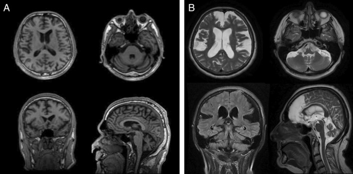
Figure 2.

MRI of the brain of two patients with pathogenic repeat expansions in FGF14. (A) Patient SCN46, who is 10 years after disease onset and shows frontal and cerebellar atrophy. (B) Patient SCN412, who is 15 years after disease onset and shows marked global brain and cerebellar atrophy.
