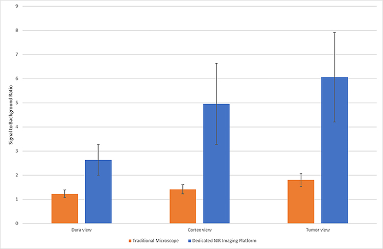
Figure 4.
Detection of NIR signal from tumor through intact dura, through cortex, or direct tumor view using both the traditional microscope with NIR add-on function and dedicated NIR imaging platform. In all three views, the dedicated platform demonstrates significantly higher signal to background ratios (t-test, p= 0.0051, 0.0026, 0.0013 respectively).

