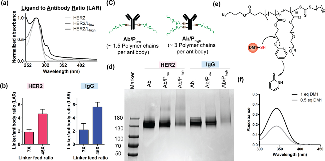
Figure 2:
Confirmation of antibody polymer conjugation and calculation of DAR. (a) UV-vis spectrum of linker conjugated antibody was used to determine the linker to antibody ratio (LAR). The ratio of absorbance at 310 nm (DBCO) and absorbance at 280 nm (antibody) was used to determine the LAR. (b) Optimized LAR of anti-HER2 and IgG (isotype control) antibodies at 7X and 45X feed ratio. (c) Representation of two antibody polymer (free polymer) conjugate system studied in this work. The number of polymer chains in each system represents the ratio of obtained number of polymers per antibody. (d) Gel electrophoresis image of antibody polymer (free polymer) conjugates for both HER2 and IgG antibodies. The tailing nature of bands for Ab/Phigh for both HER2 and IgG confirms the higher number of polymer conjugations to antibody. (e) The reaction scheme of DM1 drug conjugation to polymer chains via thiol-disulfide exchange reactions. (f) UV-vis spectrum of released byproduct (2-mercaptopyridine) from polymer chain after drug conjugation reaction. The concentration of 2-mercaptopyridine was used to determine the number drugs per polymer chains.

