Figure 3:
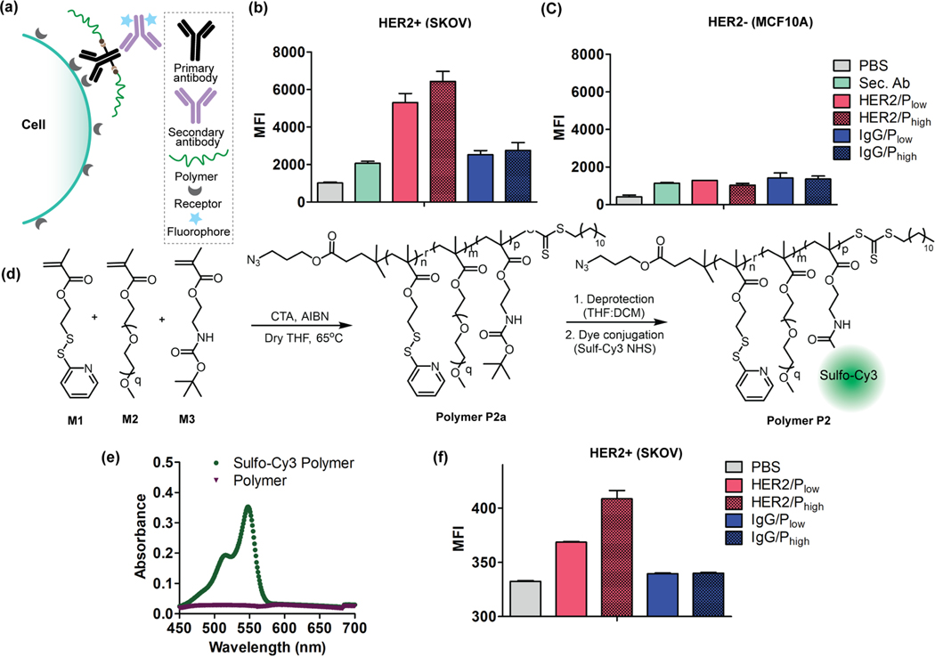
Evaluation of specificity of APCs towards HER2 receptors. (a) Schematic representation of APC binding interaction evaluation using fluorophore-labeled secondary antibody. Specificity evaluation of HER2/APCs and IgG/APCs (control) in (b) SKOV cells (HER2+) and (c) MCF10A (HER2-) cells; Sec. Ab: Secondary Antibody. (d) Synthetic scheme of polymer P2 conjugation with fluorophore (Sulfo-Cy3 NHS). (e) UV-vis spectrum of dye tagged polymer with an absorbance maximum at 548 nm confirming the successful conjugation of dye to the polymer. (f) The dye tagged polymer was conjugated to antibodies (anti-HER2 and IgG) and studies the binding specificity of APCs in SKOV3 cell (HER2+) using flow cytometry.

