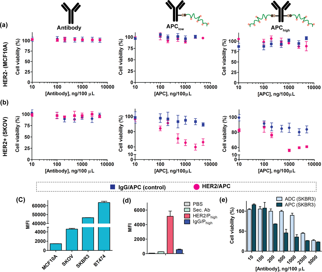
Figure 4:
Cytotoxicity evaluation of APCs in HER2+/− cells. (a) The cell viability of free antibody (both HER2 and IgG), comparative cytotoxicity of APC-Plow and APC-Phigh in MCF10A (HER2-) cells. (b) The cell viability of free antibody (both HER2 and IgG), comparative cytotoxicity of APC/Plow and APC/Phigh in SKOV3 (HER2+) cells. (c) Receptor expression quantification of HER2 in four different cell lines. (d) Specificity evaluation of APCs in SKBR3 cell using dye tagged secondary antibody. (e) Comparative cytotoxicity study of HER2/APC with Kadcyla® ADC in SKBR3 (HER2+) cell.

