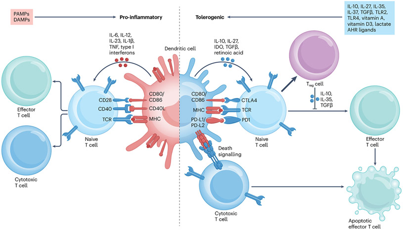
Fig. 1 ∣. Mechanisms and features in pro-inflammatory dendritic cells compared with tolerogenic dendritic cells.
Pro-inflammatory dendritic cells (DCs) can be induced via activation by pathogen-associated molecular patterns (PAMPs) and damage-associated molecular patterns (DAMPs) and upregulate the expression of surface molecules including MHC molecules, CD80 and CD86. These surface molecules, in addition to secreted pro-inflammatory cytokines, such as IL-1β, IL-6, IL-12, IL-23, tumour necrosis factor (TNF) and type I interferons, induce the differentiation of cytotoxic and effector T cells from naive T cells. Conversely, tolerogenic DCs can be induced via several mechanisms, including exposure to cytokines such as IL-10, IL-27, IL-35, IL-37 or transforming growth factor-β (TGFβ); signalling via Toll-like receptor 2 (TLR2), TLR4 or aryl hydrocarbon receptor (AHR); or exposure to molecules such as vitamin D3, vitamin A or lactate. Tolerogenic DCs express lower levels of MHC molecules, CD80 and CD86 and secrete anti-inflammatory cytokines and molecules such as IL-10, TGFβ, IL-27, indoleamine 2,3-dioxygenase (IDO) and retinoic acid. Tolerogenic DC interactions with T cells induce the differentiation and expansion of anti-inflammatory regulatory T cells (Treg cells) from naive T cells and the apoptosis of cytotoxic T cells through death receptor signalling interactions, such as between programmed cell death 1 (PD1) and PD1 ligand 1 (PD-L1) or PD-L2. CTLA4, cytotoxic T lymphocyte associated protein 4; TCR, T cell receptor.

