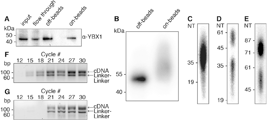
Fig. 2.
Illustration of steps during PAR-CLIP. (A) Immunoblot using antibody against the YBX1 protein. Samples from left to right: input, IP flow through, “off-beads” and “on-beads” IP elutions. (B) Autoradiograph showing the separation of radiolabeled YBX1 RNP immunoprecipitated from “off-beads” and “on-beads” procedures. Eluted RNPs are separated by SDS-PAGE and transferred to a nitrocellulose membrane. (C-E) Autoradiography of denaturing polyacrylamide gels that are used along the “off-beads” procedure to visualize and select: (C) RNA fragments extracted after Proteinase K digestion of RNPs, (D) products of 3′ adapter ligation, and (E) products of 5′ adapter ligation. (F, G) Agarose gel separation of PCR products at increasing cycle numbers for off-beads (F) and on-beads (G) procedures. For library preparation we chose 15 and 18 cycles, respectively. The expected PCR product runs at ~100 bp. Linker-linker side-products run at 71 bp.

