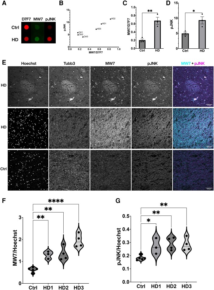
Figure 6.
Aberrant activation of JNKs and increased PRD exposure in striatal tissue from HD patients. (A) A representative dot blot of human postmortem caudate tissue (see Supplementary Table 2 for patient demographic information) showed increased immunoreactivity of MW7 and pJNK in Huntington’s disease (HD) patients, despite a moderate reduction in the total level of HTT protein as detected by D7F7 antibody. (B) Quantification of dot blots was plotted for each case (n = 3 for control and n = 3 for HD) to demonstrate a correlation between MW7/D7F7 ratio and JNK activity in HD cases, with controls clustered at the lower quadruple. (C and D) Relative immunoreactivity of MW7 (shown as ratio of MW7/D7F7) and pJNK from the same blots was quantified and plotted to show increases in HD as compared with the control samples (**P = 0.0068, *P = 0.0276, unpaired two-sample T-test). (E) Representative confocal images of the striatum indicated associated and increased immunostaining of MW7 and pJNK in HD patients compared with their controls. Tissues were counterstained with Hoechst as a nuclear marker and Tubb3 as a neuronal marker. (F and G) Quantification was performed on whole striatum images stitched from single images captured by wide-field epifluorescence imaging and showed higher MW7 and pJNK immunoreactivities in HD patients (n = 11) than in controls (n = 5) and the differences were statistically significant (****P < 0.0001, **P < 0.01, *P < 0.05 Kruskal-Wallis one-way ANOVA). HD cases were graded according to neuropathology27 (see Supplementary Table 3 for patient demographic information of cases examined in E–G).

