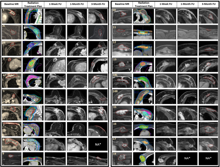
Fig 5. Baseline imaging and radiological follow-up at different time points.
The figure details the radiation treatment plan and magnetic resonance imaging of the target tumour at baseline, 1 week, 1 month, and 3 months posttreatment. Patients were assigned numbers from 1 to 18, and for those with bilateral tumours, the left and right sides were denoted by “L” and “R,” respectively (i.e., “2L” and “2R”). The dotted red line represents the target tumour at baseline and the residual tumour or replacement fibrosis at 3 months follow-up imaging. In the radiation treatment plan, the 105% isodose line was represented in yellow, the 100% in red, the 95% in green, the 80% in dark blue, and the 50% in light blue. *Two patients died before the 3-month follow-up, represented by NA*. FU, follow-up; MR, magnetic resonance imaging; NA, not available; L, left; R, right.

