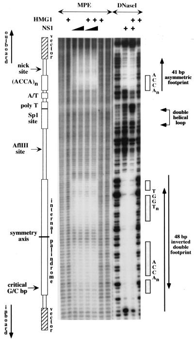
FIG. 6.
A schematic representation of the origin is aligned down the left-hand side of autoradiographs from denaturing polyacrylamide gels displaying hydroxyl radical (MPE) and DNase I cleavage profiles obtained with naked 32P-, 3′-end-labeled DNA or following its incubation with NS1 and/or HMG1 as indicated. Each lane displays products from the upper strand, containing the nick site. The positions of the ACCA tetranucleotide motifs that make up the NS1 binding sites are indicated down the right-hand side, together with a region of overcutting and undercutting, obtained with DNase I in the presence of both NS1 and HMG1, that reflects the formation of a double helical DNA loop.

